
Figure 3.
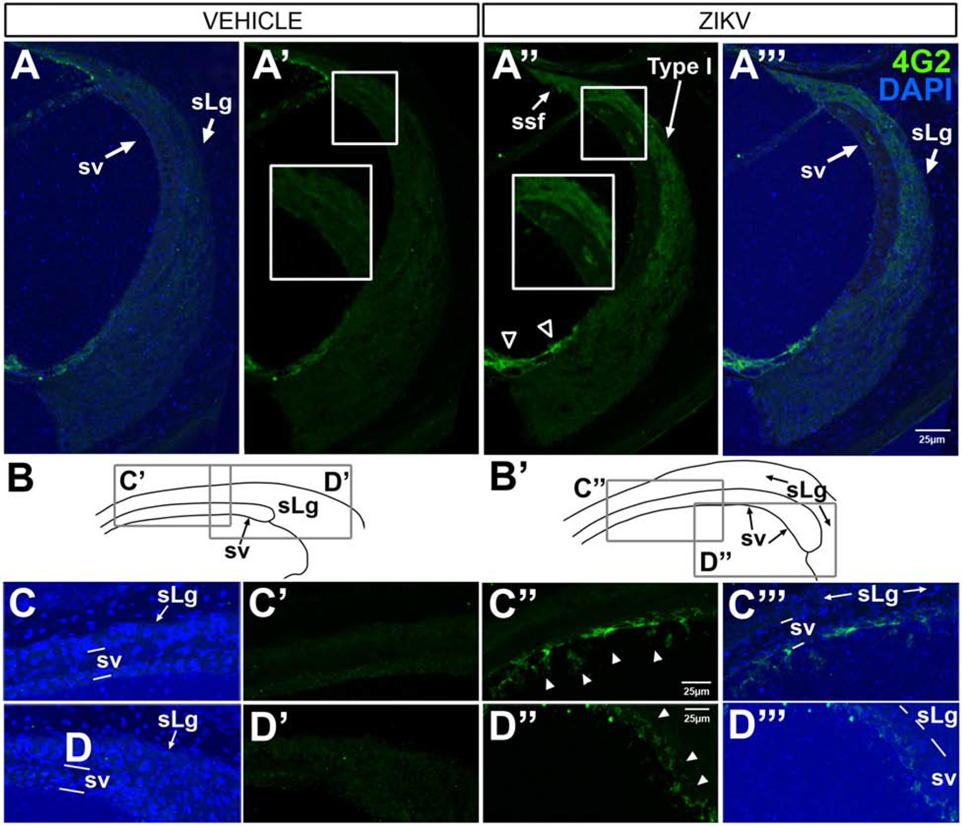
4G2 protein is localized in the lateral wall of the cochlea in Ifnar1−/− ZIKV infected mice. Through the midturn of the cochlea, 4G2 is not seen in the spiral ligament or stria vascularis in vehicle-treated cases (A’; DAPI-counterstained, A), but 4G2 is localized in the lateral support cells (open arrowheads in A”, A”’), the spiral ligament (sLg), suprastrial (ssf) and Type I fibrocytes (A”, A”’) and cells within the laminae of the stria vascularis (A”, A”’) after ZIKV infection. The small boxed regions (A’ and A”) showing regions of higher magnification views in the larger boxes (A and A”) reveal 4G2 localization in the stria vascularis and spiral ligament after ZIKV infection (A”) and not after vehicle injection (A’). The apical turn of the cochlea contains 4G2-positive cells (arrowheads C”, D”, C”’, D”’) with different cellular morphologies dependent on location. Line drawing schematics of vehicle controls (B) and ZIKV infected (B’) indicate regions shown at higher magnification C-D”’). DAPI counterstains are shown (A, C, D, A”’, C”’,D”’). Scale bar for A - A” in A”’, scale bar for C, C’ and C”’ in C” and scale bar for D. D’ and D”’ in D”.