
Figure 5.
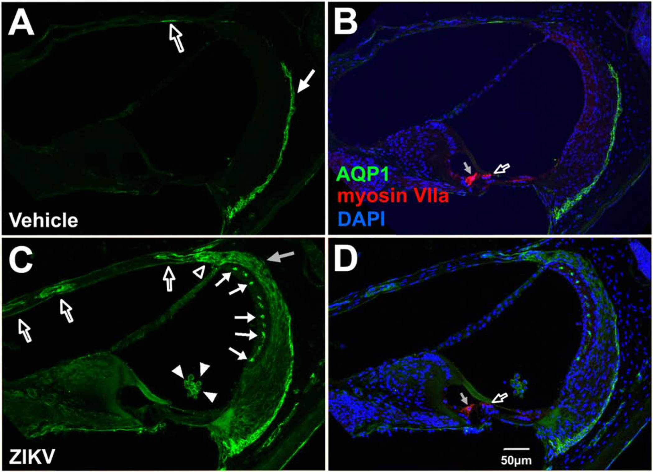
Aquaporin-1 localization in the cochlea, scala media and lateral wall of the cochlea in Ifnar1−/− ZIKV infected mice. Vehicle-injected mice show baseline AQP1 localization in type III fibrocytes of the spiral ligament (arrow in A; B) and a low level of protein localization in the endosteum of the scala vestibuli (open arrow in A; B). Postnatal ZKV infection shows upregulation of AQP1 in suprastrial fibrocytes (open arrowhead, C), Type I fibrocytes in the spiral ligament (gray arrow, C), the intermediate region of the stria vascularis (closed arrows, D) and in structures with a vascular appearance in the osseous spiral lamina above the scala vestibuli (open arrows, C). AQP1 is also localized to cells in the scala media (closed arrowheads in C; D) whose nuclei are stained with DAPI (D). Myosin VIIa of inner (gray arrow, B and D) and outer (open arrow, B and D) hairs cells are not double labeled with AQP1 with vehicle treatment (B) or ZIKV infection (D). DAPI counterstains are shown (B, D). Scale bar for A-C in D.