
Figure 6.
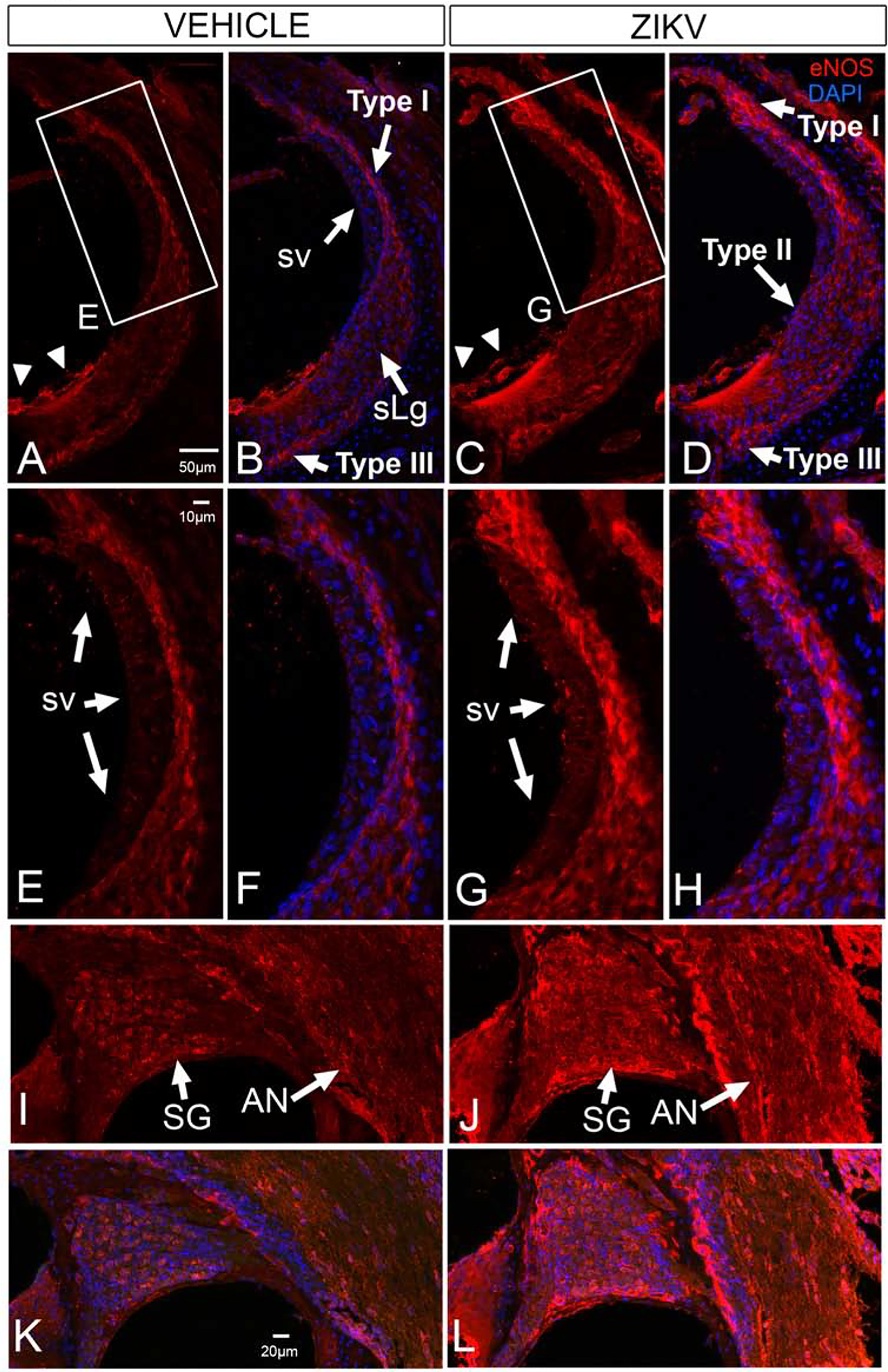
eNOS is upregulated the cochlear lateral wall, spiral ganglion and auditory nerve of Ifnar1−/− ZIKV infected mice. Baseline eNOS localization is seen in Type I and Type III fibrocytes of the spiral ligament (sLg, A, B, E, F), stria vascularis (sv, A, B, E, F), in lateral support cells (arrowheads in A; B), neurons of the spiral ganglion (SG, I, K) and the auditory nerve (AN, I, K) in vehicle injected mice. ZKV infection shows upregulation of eNOS in the Type I, Type II and Type III fibrocytes in the spiral ligament and stria vascularis (C, D, G, H) where immunoreactivity appears as larger aggregates. eNOS is also upregulated in spiral ganglion neurons in a nonneuronal pattern and in the auditory nerve (J, L). eNOS is not upregulated in the cochlear epithelium after ZIKV infection (cf C, D to A, B). DAPI counterstains are shown (B, D, F, H, K, L). Boxed regions in A and C shown in E and G, respectively. Scale bar for B-D in A. Scale bar for F-H in E. Scale bar for I-L in K.