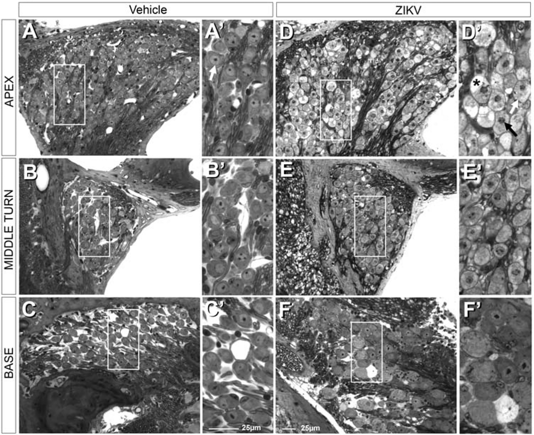
Figure 9.

Postnatal ZIKV infection of Ifnar1−/− mice produces graded damage in spiral ganglion neurons (boxed regions in A-C indicate regions shown at higher magnification in A’-C’, respectively). Following postnatal vehicle treatment, immune compromised mice maintain normal neuronal morphology of spiral ganglion neurons throughout the cochlear spiral (A-C). This includes normal cytoplasm and nuclei with prominent nucleoli (white arrow, A’). Postnatal ZIKV infection results in apparently swollen axons and fibers in the auditory nerve and surrounding the spiral ganglion neurons (D-F). Boxed regions in D-F shown at higher magnification in D’-F’, respectively. Spiral ganglion neurons at the apex (D and D’) have mottled cytoplasm (white arrow), vacuoles (asterisk) and condensed nuclei (black arrow, D’); nucleoli were not readily detectable. In the middle turn of the cochlea, fewer spiral ganglion neurons have mottled cytoplasm (E and E’). Even fewer spiral ganglion neurons with mottled cytoplasm are seen at the base of the cochlea (F and F’). While rare, some ganglion cell degeneration is still observed in basal turns (F’). Some spiral ganglion neurons at the base of the cochlea show relatively normal neuronal morphology with euchromatic nuclei, prominent nucleoli and uniformly stained cytoplasm, but are larger in size than vehicle treated spiral ganglion neurons at the base of the cochlea (cf. F to C and F’ to C’).
