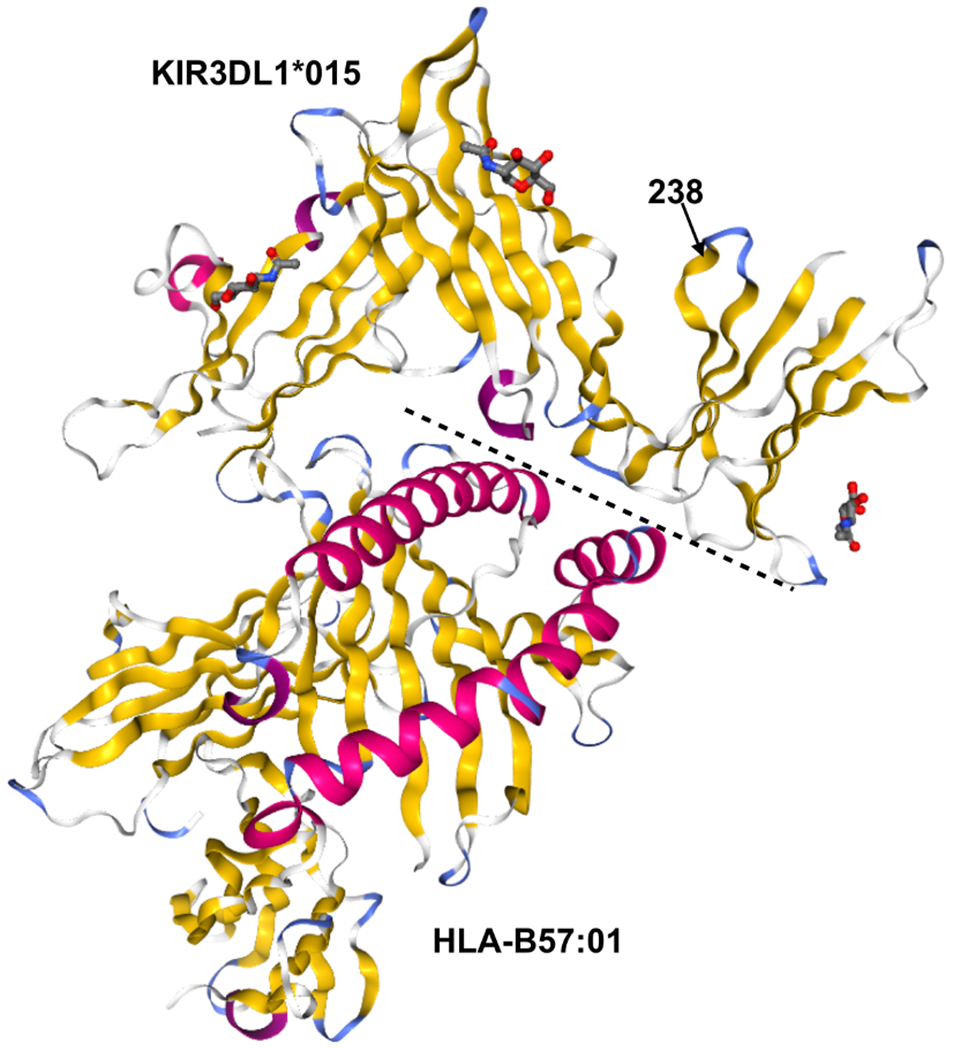
Figure 4. KIR3DL1*015 and KIR3DL1*002 Differ by A Single Amino Acid at Position 238.

Crystal structures of KIR3DL1*015 (top) complexed with its ligand HLA-B*57:01 (bottom). Secondary structures are shown by color: alpha helices (pink) and beta sheets (yellow). The dashed line indicates the binding interface between KIR3DL1*015 and HLA-B*57:01. Position 238 is indicated by an arrow. Protein Data Bank accession no. 5B39 (41).
