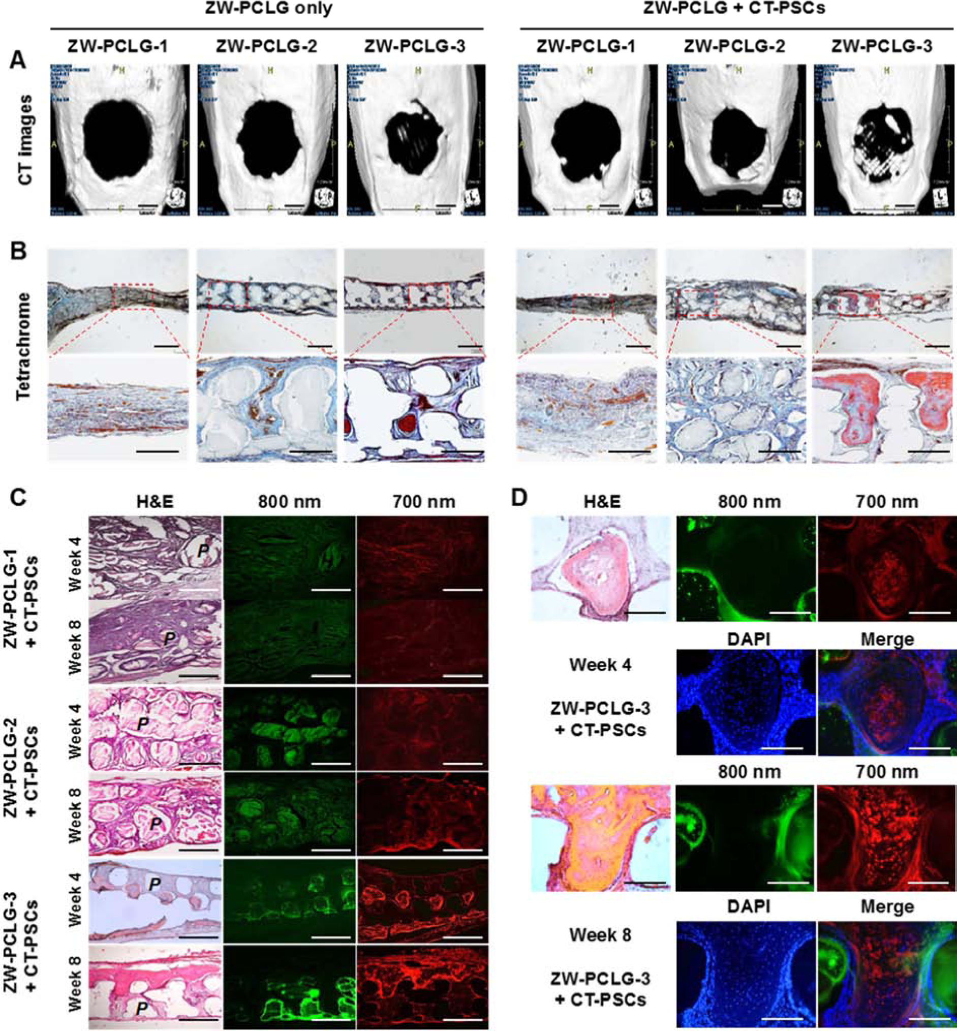
Fig. 6.

CT and histological examinations of the CT-PSCs and PCLG scaffolds. New bone formation in the calvarial bone defect was examined by (A) CT (scale bar = 2 mm) and (B) modified Tetrachrome staining (scale bar = 1 mm). (C) The remained PCLG scaffolds and hPSCs were detected by H&E staining and NIR imaging (P = PCLG). Green pseudo color at 800 nm indicates the remained PCLG scaffold, and red pseudo color at 700 nm indicates hPSCs. (D) High power images to confirm hPSCs within bone matrix in ZW-PCLG-3 + CT-PSCs. The blue color indicates DAPI-stained nuclei. Scale bar = 500 μm.
