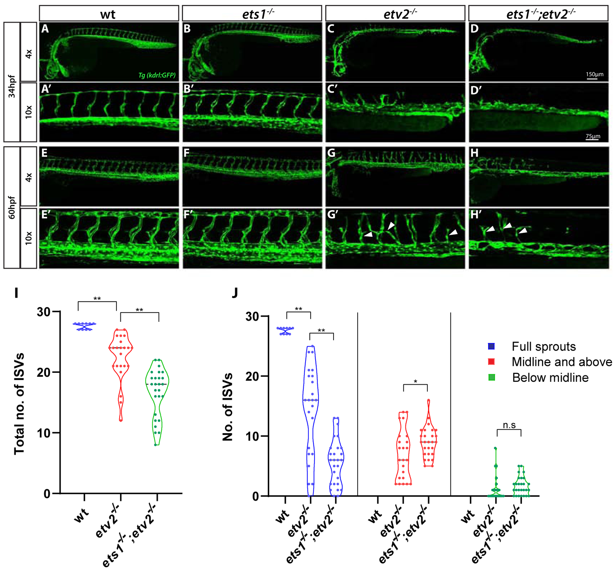
Figure 1. Loss of Ets1 function exacerbates vascular defects in etv2−/−embryos.

(A-H’) Confocal images of Tg(kdrl:GFP) expression in wt, ets1−/−, etv2−/−and ets1−/−; etv2−/−embryos at 34 hpf and 60 hpf using 4× and 10× objectives. ets1−/−embryos display normal vascular patterning (B-B’, F-F’), similar to wild type embryos (A-A’,E-E’). ISVs begin to emerge in etv2−/−embryos at ~34 hpf (C’) but are absent in ets1−/−; etv2−/− double mutant embryos (D’). At 60 hpf, ets1−/−; etv2−/−embryos have fewer ISVs and less recovery in axial vasculature than etv2−/− embryos (G-H’). (I-J) Quantitative analysis of total number of ISVs and ISV height at 60 hpf. Note the reduction in number of ISVs (I) and full sprouts (J) in ets1−/−; etv2−/−compared to etv2−/− embryos. *P<0.05, ** P <0.01; n.s – not significant. Horizontal bars within violin plots represent median. Arrowheads indicate mis-patterned and stunted ISVs.
