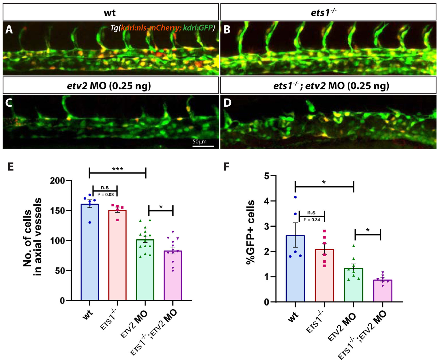
Figure 3. Loss of Ets1 function impairs vasculogenesis in Etv2-deficient embryos.

(A-D) Confocal micrographs of 28 hpf Tg(kdrl:GFP; kdrl:nls-mCherry) wild-type, ets1−/−, etv2 MO (0.25ng) and ets1−/−; etv2 MO (0.25 ng) zebrafish embryos (trunk region). (E) Quantitative analysis of endothelial cell number in the axial vessels of embryos in A-D by cell counts in Imaris. (F) FACS analysis of GFP+ endothelial cells in 28 hpf embryos. Note the decrease in endothelial cell number between etv2 MO and ets1−/−; etv2 MO embryos (E,F). Lateral views; anterior is to the left. *P<0.05, *** P<0.001; n.s – not significant; error bars represent ± SEM.
