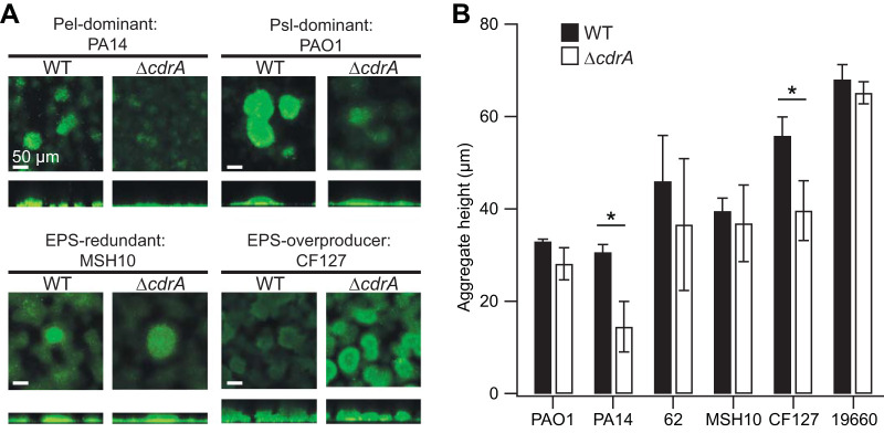
FIG 3.
CdrA promotes biofilm aggregation in flow cells. (A) Representative flow cell biofilms of several isolates stained with Syto9 and imaged using confocal laser scanning microscopy (CLSM). In general, the ΔcdrA mutants formed shorter biofilm aggregates than their wild-type (WT) counterparts. (B) The difference in aggregate height was quantified. Error bars represent the standard deviations for 3 or 4 biological replicates. The values for each biological replicate were obtained from measurements of 4 to 8 aggregates per flow cell (*, P < 0.05 [t test]).

