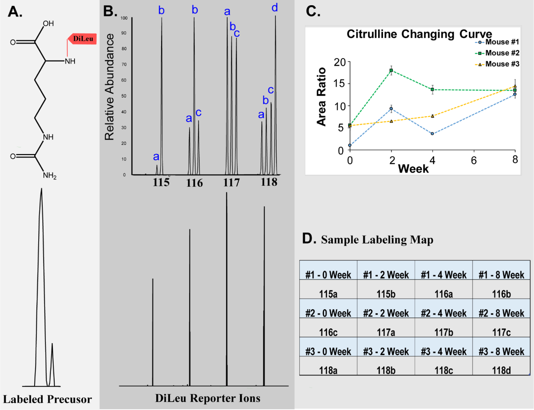
Figure 1. Details of 12-plex DiLeu labeling.

A: Precursor ion of DiLeu-labeled citrulline; B: An MS2 spectrum of the 12-plex DiLeu-labeled citrulline acquired in the Orbitrap at 60 K resolving power. Low m/z region showing distinct DiLeu reporter channels (bottom) and after zooming in, twelve distinct reporter ion peaks are present (bottom); C: Citrulline changing trends from different time points of three biological replicates. D: Sample labeling map showing the 12-plex DiLeu tags and time point (randomized)
