Figure 7.
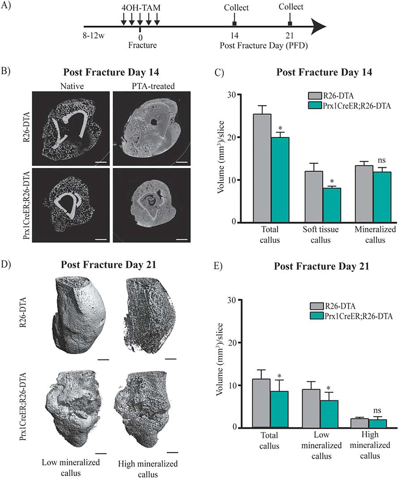
Defective fracture healing in Prx1CreER;R26-DTA mice.
(A) Schematic representation of 4OH-TAM injections in fractured Prx1CreER;R26-DTA and R26-DTA, control. (B) Representative μCT images of control and Prx1CreER;R26-DTA mice at 14 days after fracture, with or without PTA staining (10 days). Scale bars= 1mm. (C) Volumes of total callus, soft tissue and mineralized callus are normalized to the number of slices (mm3/slice) comprising the callus (see Material and Methods section for more details). Data are reported as mean ± SD of duplicate repeats from 5 separate samples. *, p < 0.05, compared to Control by unpaired two-tail t-test. (D) Representative three-dimensional μCT images of control and Prx1CreER;R26-DTA and R26-DTA mice at 21 days after fracture. Scale bars= 1mm. (E) Volumes of total mineralized callus, low mineralized callus and high mineralized callus are normalized to the number of slices (mm3/slice) comprising the callus. Data are reported as mean ± SD of duplicate repeats from 5 separate samples. *, p < 0.05, compared to Control by unpaired two-tail t-test.

