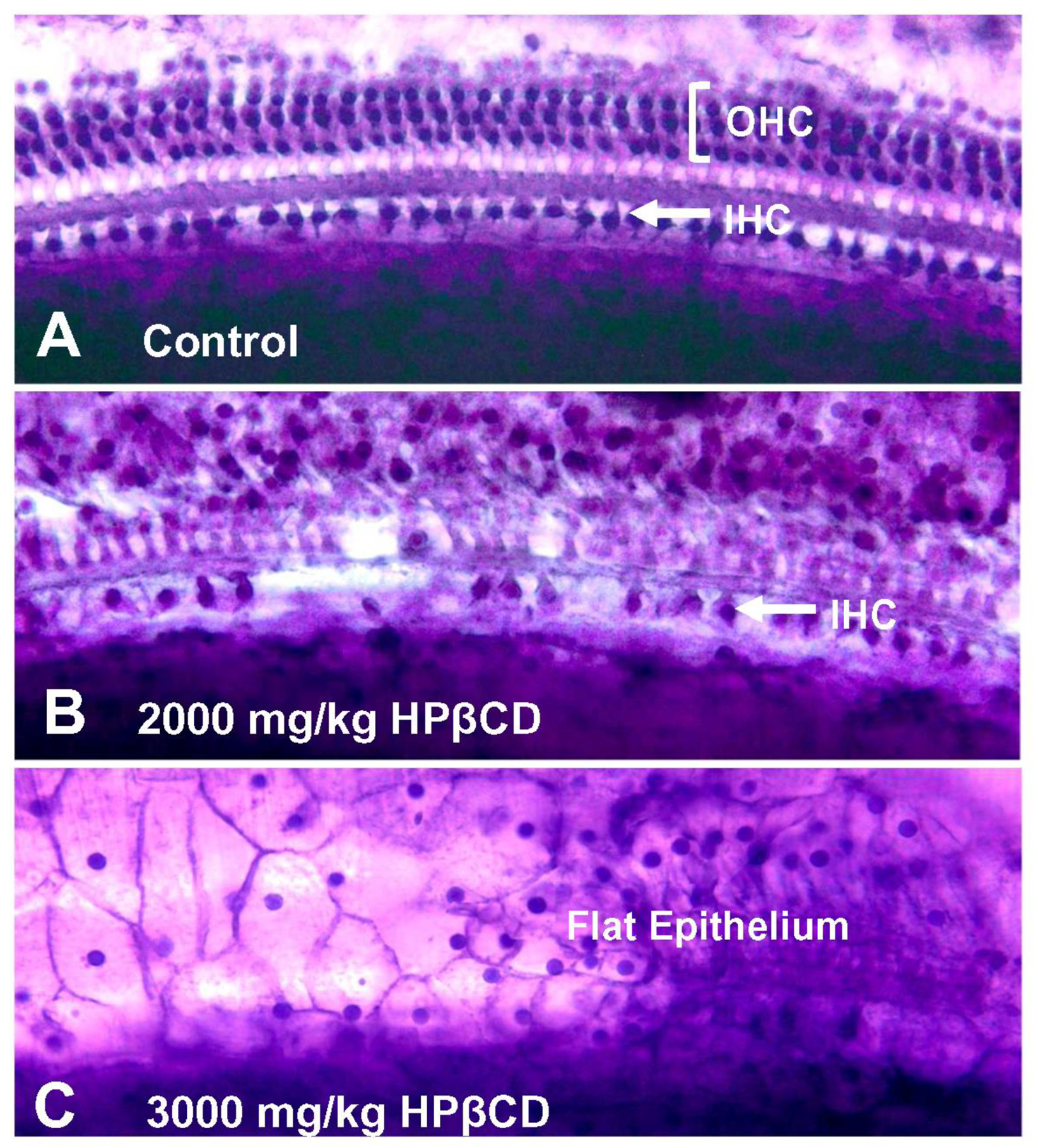
Figure 10:

Representative photomicrographs of cochlear surface preparations stained with Harris’ hematoxylin solution. (A) Surface preparation from saline control showing nuclei in three orderly rows of outer hair cells (OHC) and a single row of inner hair cells (IHC). (B) Photomicrograph from base of cochlea of rat treated with 2000 mg/kg of HPβCD showing partial loss of IHC and absence of OHC. (C) Photomicrograph from cochlea of rat treated with 3000 mg/kg of HPβCD showing flat epithelium lined with cuboidal cell profiles and absence of any IHC or OHC.
