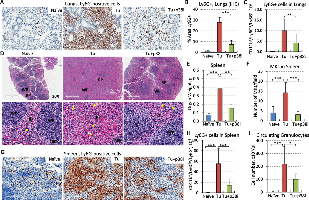
Figure 3. Blockade of p38 reduces accumulation of MDSCs in the lungs and the expansion of PMN-MDSCs and megakaryocytes in the spleen of tumor-bearing mice.
(A) Lung tissues from tumor-free (naïve), 4T1-Luc tumor-bearing (Tu) and tumor-bearing mice treated with p38i were stained for Ly6G, a marker of PMN-MDSCs, bar=200μm. (B) Quantification of Ly6G+ cells in the lung sections, 3 fields per section, 3 mice per group. (C) Flow cytometry analysis of CD11b+Ly6CloLy6G+ cells in whole-lung tissues, 3 mice per group. (D) H&E-stained spleen tissues at 20x magnification (top row, bar=1mm), bottom at 200x, bar=100μm. Yellow arrows show megakaryocytes; WP, white pulp; RP, red pulp. Note an increase in the RP and spleen size in tumor-bearing mice compared to tumor-free mice, and their reduction by p38i/Ralimetinib. (E) Spleen weights were measured at day 14 of the study, n=5. (F) Evaluation of megakaryocytes (MK) in the spleen of tumor-free (naïve), tumor-bearing (Tu), and tumor-bearing treated with p38i (Tu+p38i). (G) Spleen sections stained for Ly6G, bar=100μm. (H) Flow cytometry data for CD11b+Ly6CloLy6G+ cells in whole-spleen tissue from 3 mice per group. (I) Quantification of granulocytes in peripheral blood using complete blood count (CBC) assays. Comparisons were done with material from naïve mice and mice with comparably sized tumors, and statistical significance was determined using the two-tailed unpaired t-test (*P<0.05; **P<0.01; ***P<0.001).

