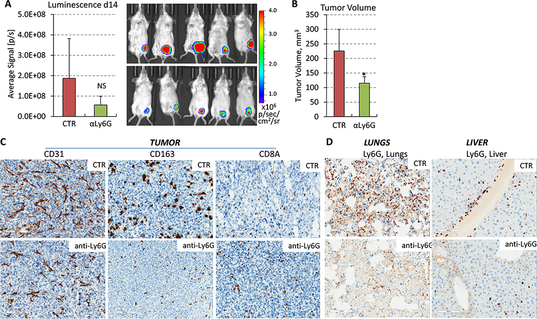
Figure 5. Depletion of PMN-MDSCs by anti-Ly6G antibody impedes the pre-metastatic niche.
(A) Panels show bioluminescence images and their quantification in live animals at day 14. 4T1-Luc mammary carcinoma cells (200,000) were orthotopically implanted in female BALB/c 6-week old mice, 7 mice/group. The next day, mice were administrated i.p. injections of vehicle-control (saline) or antibody to Ly6G scheduled on every other day. (B) Tumor volume at day 14. Statistical significance was determined using the two-tailed unpaired t-test (*P<0.05). (C) Blood vessels (CD31), tumor-associated macrophages (CD163) and tumor-infiltrating T cells (CD8A) were visualized in tumor sections from comparable-size tumors (day 14) treated with anti-Ly6G or isotype-control (CTR) antibody, bar=100μm. (D) Panels show Ly6G stained sections of the lungs and liver of mice treated with anti-Ly6G or isotype-control (CTR) antibody, bar=100μm.

