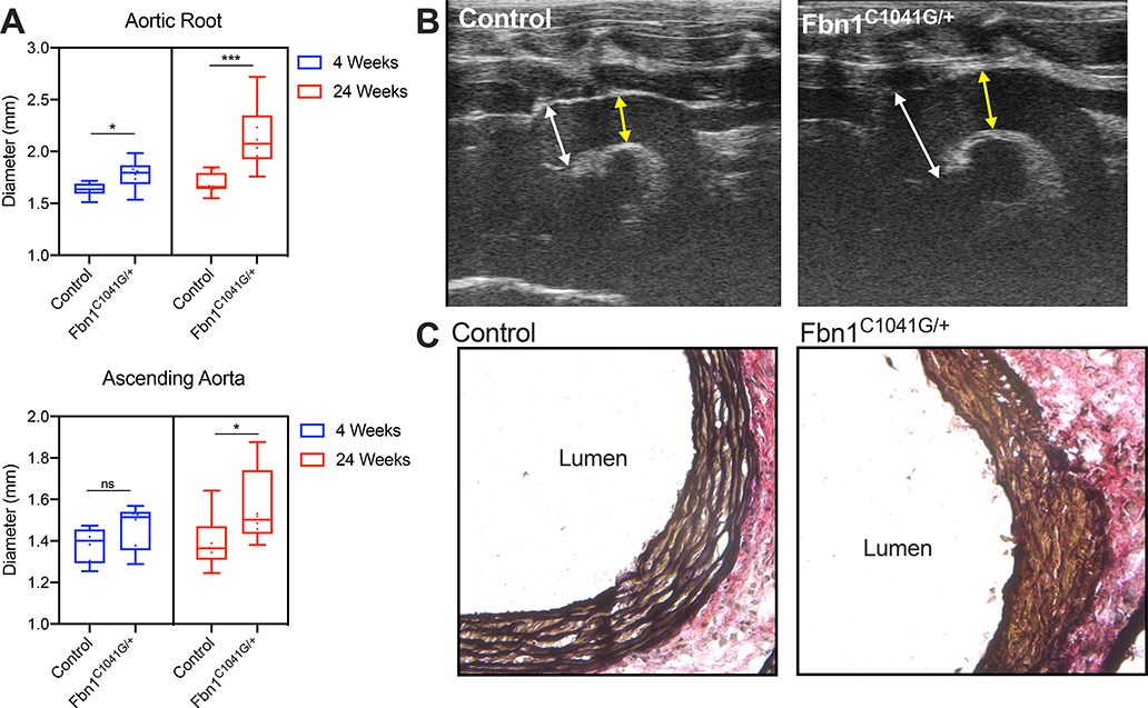
Figure 1:
Progressive aortopathy in Fbn1C1041G/+ mice. A Long axis echocardiographic aortic root and ascending aorta measurements in 4-week (n=6 mice per genotype) and 24-week old (n=8 per genotype) control and Fbn1C1041G/+ mouse cohorts. B Representative 24-week transthoracic echocardiographic images demonstrating severe dilatation of the aortic root in Fbn1C1041G/+ mice. White arrows depict sinus of Valsalva measurement. Yellow arrow depicts ascending aorta measurement. C Representative elastic Van Gieson (EVG) stain demonstrating severe elastin fiber fragmentation in Fbn1C1041G/+ mice. Box and whisker plots display 95% confidence interval (box), mean (line) and range (whiskers). ***p<0.001, *p<0.05 (Mann-Whitney U non-parametric test).

