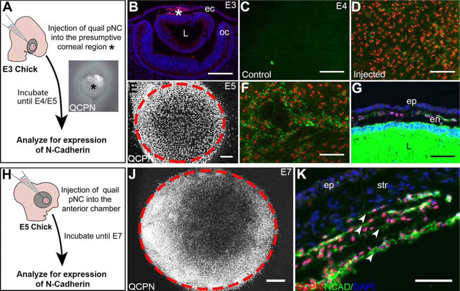
Figure 2. pNC injected directly into the presumptive corneal region at different timepoints of development differentiate into corneal endothelial cells.

(A) Schematic overview of the injection of pNC into E3 presumptive corneal region (asterisk) and further experimental procedures. (B) cross-section through E3 eye, showing QCPN-positive pNC (asterisk) in the presumptive corneal region between the ectoderm and lens. (C) Wholemount immunostaining of an E4 chick eye showing no NCAD expression in the presumptive corneal region prior to the pNC migration comparing to (D) Wholemount immunostaining of an E4 chick eye showing the NCAD expression surrounding some of the QCPN-positive cells 1 day post-injection at E3. (E) Wholemount immunostaining of an E5 chick eye showing the localization QCPN-positive pNC in the cornea (delineated by the dotted circle) and surrounding region 2 days post injection at E3. (F) Injected pNC stain positive for NCAD at E5. (G) Cross-section of an E5 eye showing that the injected pNC form an NCAD-positive monolayered corneal endothelium. (H) Schematic overview of the injection of the pNC into E5 presumptive cornea and further experimental procedures. (J) Wholemount immunostaining of an E7 chick eye showing the localization QCPN-positive pNC in the cornea (delineated by the cells and dotted circle) following 2 days post injection at E5. (K) Cross-section through E7 eye, showing that injected cells form multiple layers of QCPN- and NCAD-positive cells (arrowheads). ec, ectoderm; en, corneal endothelium; ep, corneal epithelium; L, lens; oc, optic cup; str, corneal stroma. Scale bars represent 100 μm (B,E); 50 μm (C, D, F, G, J, K).
