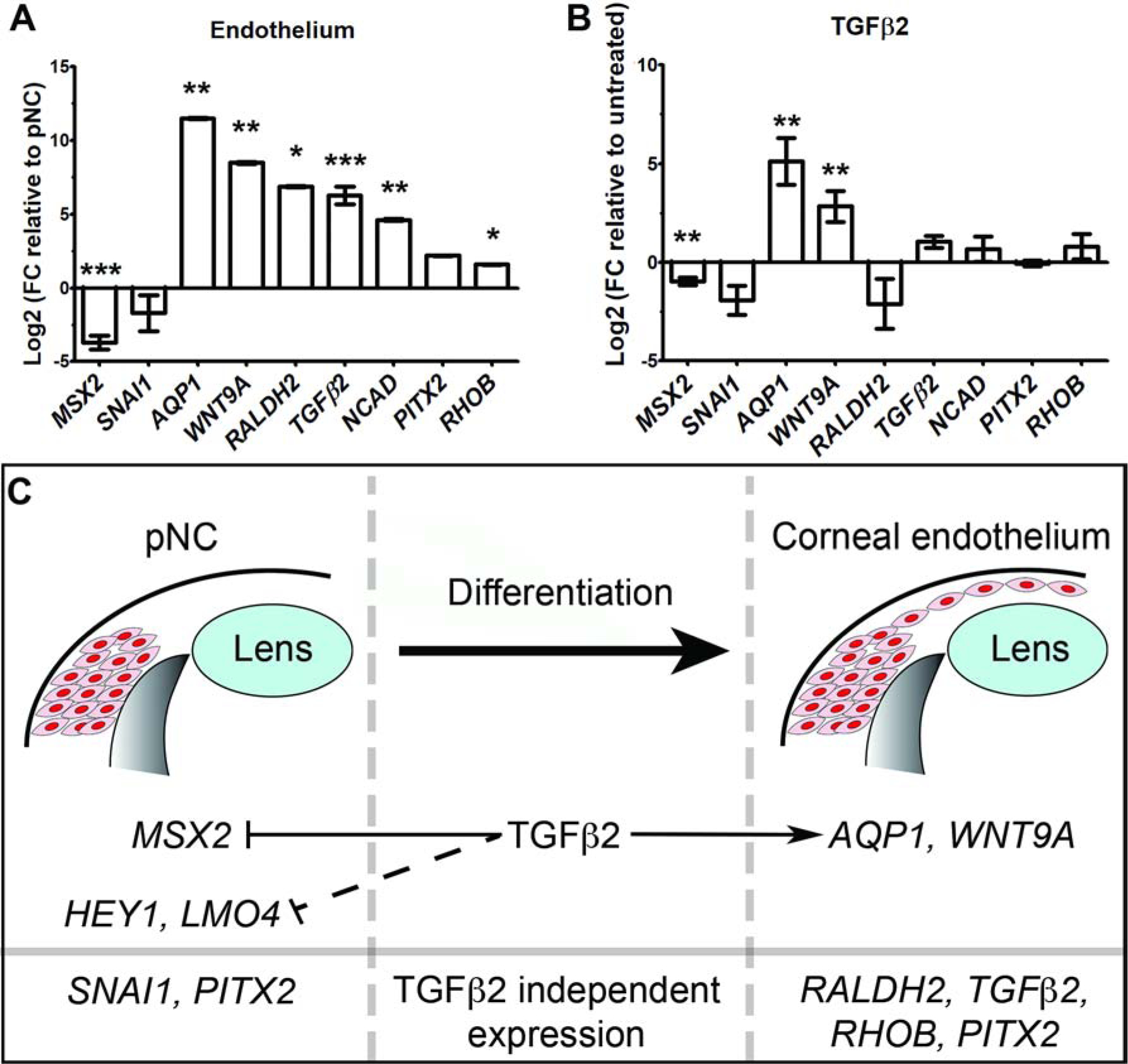
Figure 6. Molecular mechanisms of pNC differentiation into the corneal endothelium.

(A-B) Comparative qPCR analysis of pNC and endothelial mRNA expression under different conditions. GAPDH was used as reference gene. Independent sample replicates n=3. *P<0.05, ** P<0.01, *** P<0.001 (Welch`s two-tailed t-test). Data represented as mean ± s.d. (A) Fold change (FC) of corneal endothelial genes compared to E2.5 pNC (baseline). (B) Fold change in gene expression of TGFβ2-treated pNC compared to untreated control culture (baseline). (C) Schematic representation of the regulation of pNC and corneal endothelial genes by TGFβ2 signaling.
