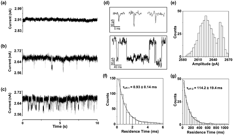
Figure 2.

Detection of HIV-1 protease in a PET nanopore. Typical 10-s single channel recording trace segments of a) 100 ng/mL HIV-1 protease in the unmodified PET nanopore, b) 10 ng/mL and c) 100 ng/mL HIV-1 protease in the amine-modified PET nanopore; d) selective individual short-lived and long-lived events; and the corresponding event (e) amplitude histogram and residence time histograms of (f) short-lived and (g) long-lived events of Figure 2c. The experiments were performed at +800 mV in a solution comprising 1 M KCl and 10 mM Tris (pH 7.5). An uninterrupted 2-min single-channel recording trace of 100 ng/mL HIV-1 PR in the modified PET nanopore was displayed in Figure S4.
