
Fig. 3. Mode of intercellular mitochondrial transfer.
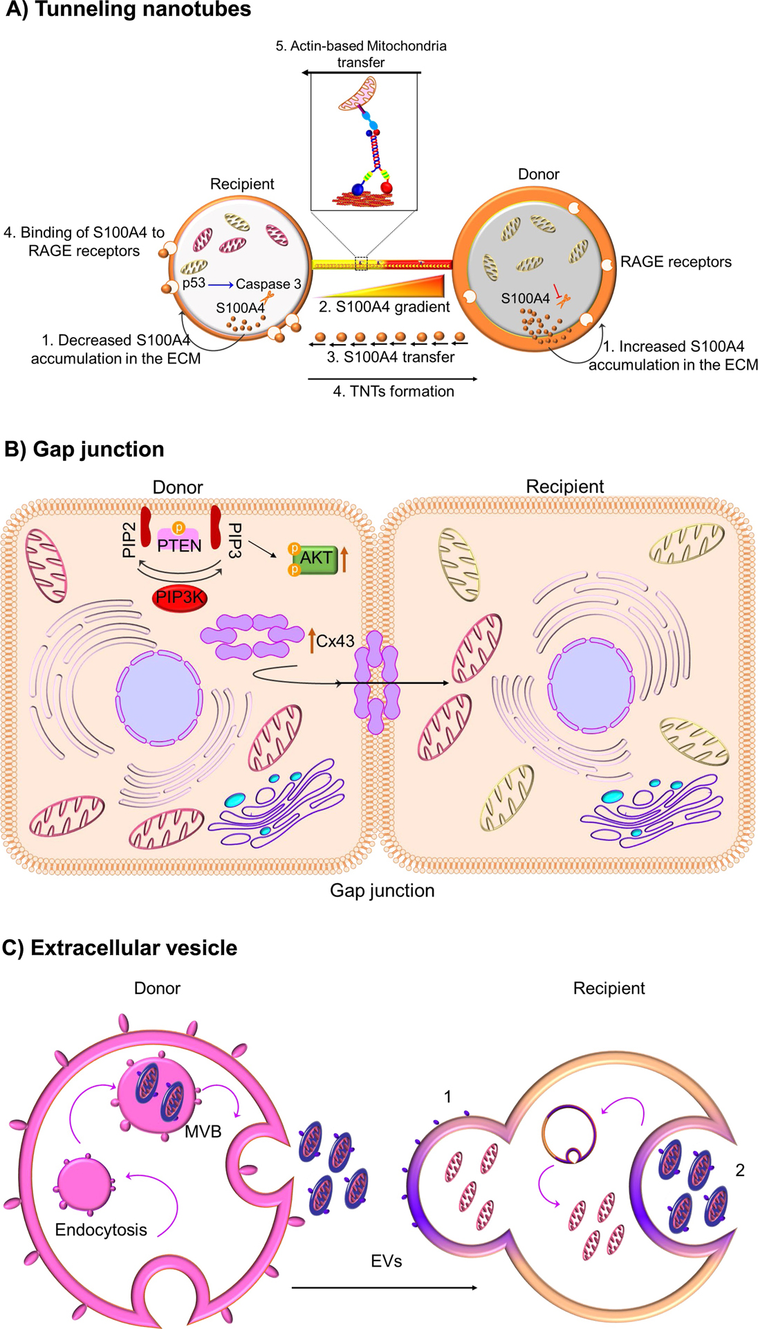
(A) Tunneling nanotubes.P53-mediated activation of caspase 3 cleaves S100A4, creating a gradient of low levels of S100A4 in initiating or recipient cells (injured cells) towards a higher concentration in target or donor cells (healthy cells). It is suggested that RAGE in recipient cells act as the putative receptor for the high concentration of S100A4 in donor cells and this co-ordination of S100A4 and RAGE guides TNT direction. Mitochondria transfer through nanotunnels is believed to be facilitated by Actin/Miro-based transport machinery. (B) Gap junction. Connexin, specifically connexin 43 oligomerizes to form a channel at the gap junction which facilitates the transfer of mitochondria. Mechanistically, it was shown that ROS-induced oxidative stress regulates the opening of connexin channels in a system mediated by phosphoinositide 3-kinase (PI3K). (C) Extracellular vesicle. The multivesicular bodies (MVB) release extracellular vesicles (EVs) from the donor cell. These released EVs can fuse with recipient cells (1) or engulfed (2) to release the EVs’ content