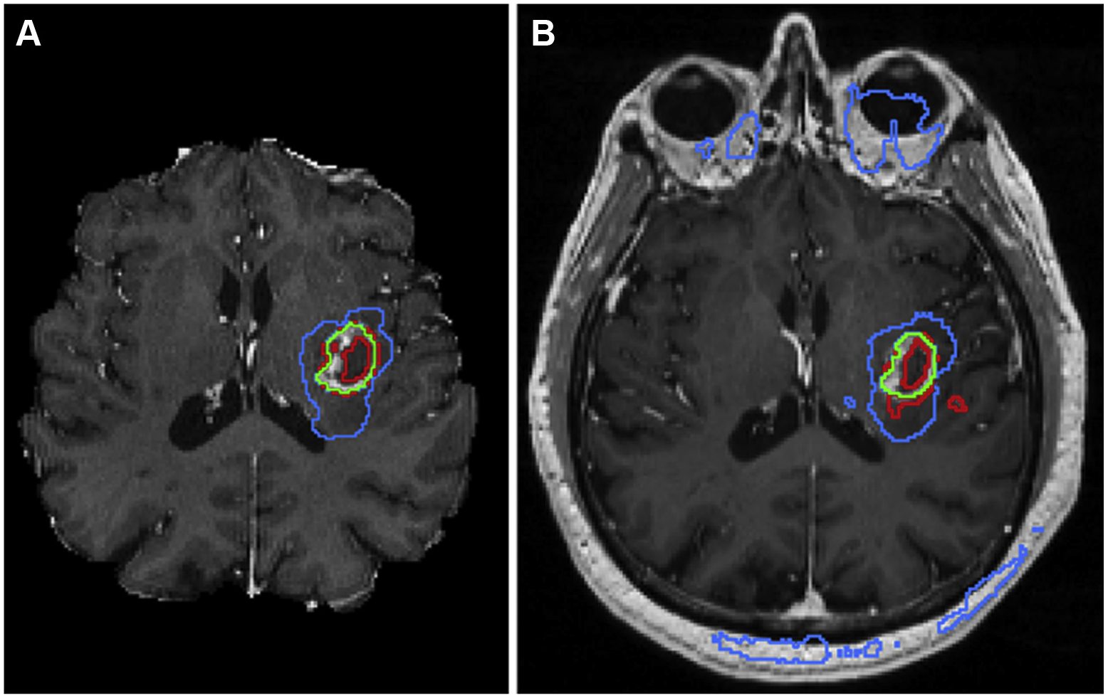
Fig 2.

Implementation of one of the top-performing segmentation algorithms from the 2017 Medical Image Computing and Computer Assisted Intervention Brain Tumor Segmentation Challenge shows excellent results (A) when data are preprocessed using skull-stripping and registration algorithms before inputting into a segmentation neural network; however, when the algorithm is deployed on a clinical MRI data set, the resulting segmentation contains many errors (B). Here, image segments identified as enhancing tumor are outlined in red, regions of enhancing tumor with any central nonenhancing components are shown in green, and regions of tumor with surrounding edema are shown in blue.
