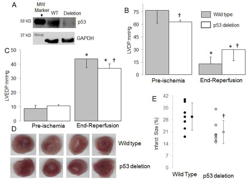
Figure 1. Deletion of p53 decreases cardiac injury in mouse hearts following ischemia-reperfusion.
The content of p53 was decreased in cardiomyocyte specific deletion mice compared to wild type mice (WT) (Panel A). Mouse hearts were buffer perfused and exposed to 25 min. ischemia and 30 min. reperfusion. Cardiac contractility [reflected by left ventricular developed pressure (LVDP, mmHg)] was decreased following 60 min reperfusion (Panel B), whereas deletion of p53 improved contractile function compared to wild type mice (Panel B). Deletion of p53 also improved diastolic relaxation in hearts following 60 min reperfusion compared to wild type (Panel C). Representative pictures for infarct size measurement from wile type and p53 deletion mice are shown in Panel D. The infarct size was decreased in p53 deletion mice compared to wild type (Panel E). Mean ± SD. * p<0.05 vs. corresponding pre-ischemia. † p<0.05 vs. corresponding wild type hearts. n=7 in each group.

