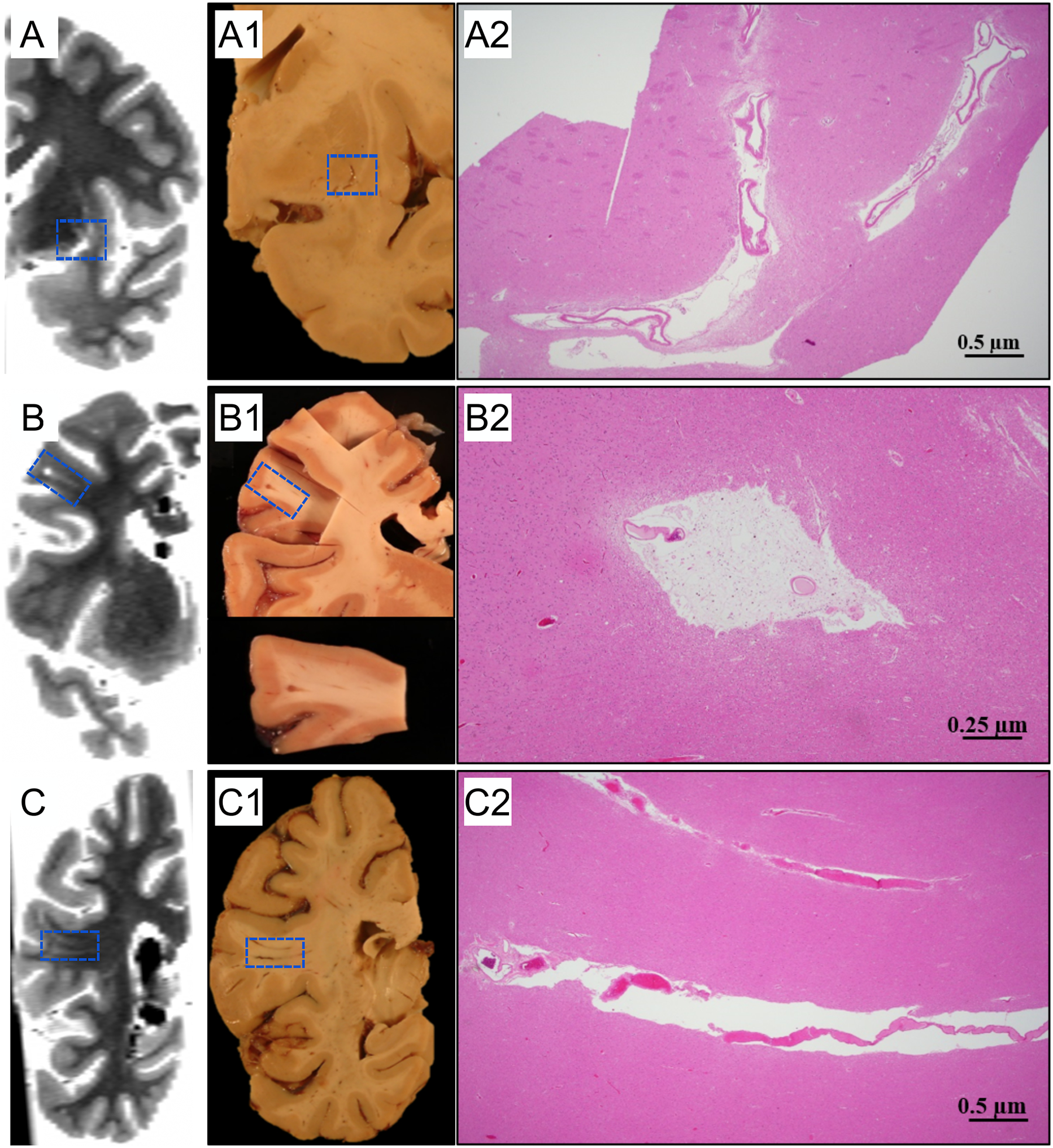
Figure 2.

Examples of histopathologic validation of EPVS. Three participants with mild (A), moderate (B), and severe (C) EPVS burden are shown. The first column (A,B,C) shows ex-vivo MR images that have been reformatted to match the plane shown in the pictures of the tissue slabs (A1,B1,C1). Histological findings show dilated PVS (A2, B2, C2), tissue rarefaction around blood vessels (A2), hemosiderin-laden perivascular macrophages, and severe hyaline thickening of small arterioles (B2).
