Figure 2.

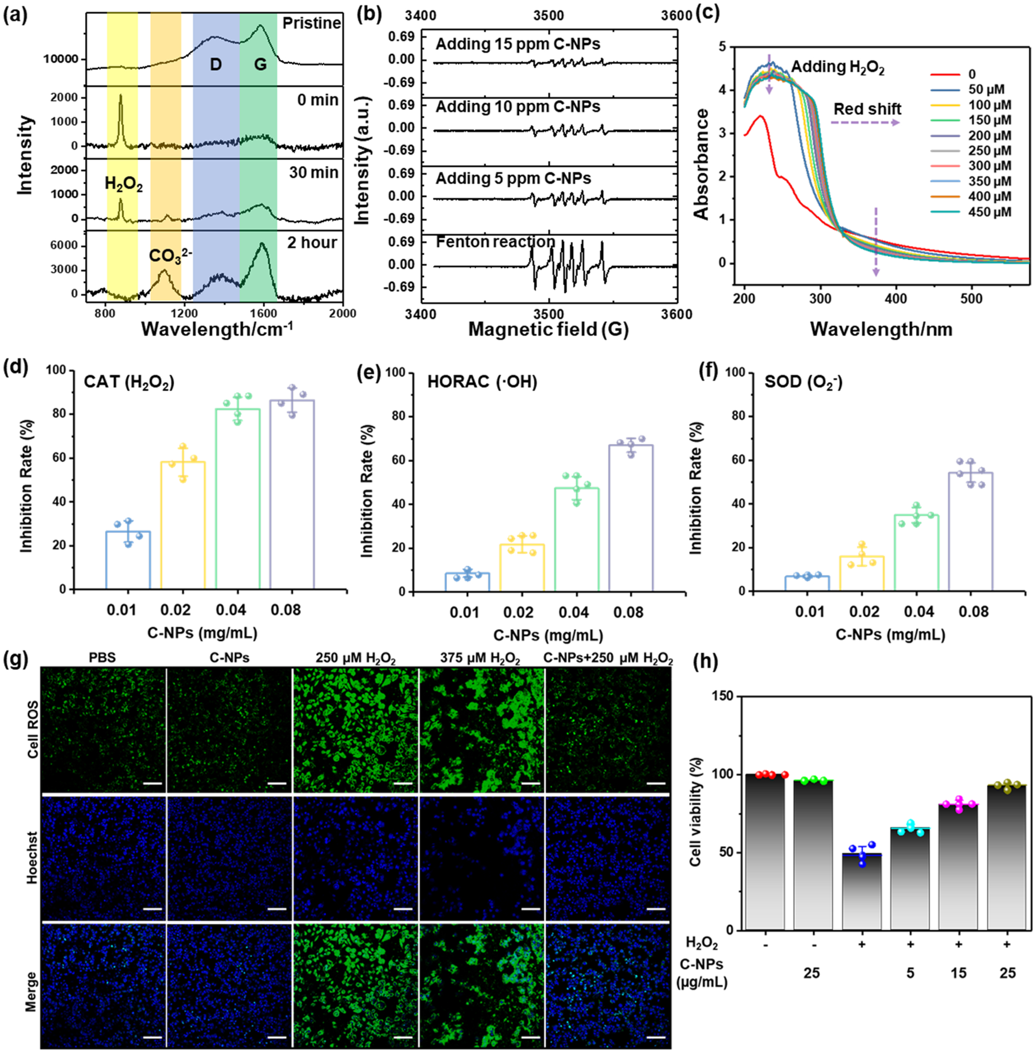
In vitro ROS scavenging capability of C-NPs. (a) In situ Raman spectra of C-NPs reacting with H2O2 at varied time points (0, 30 min, and 2 h). (b) ESR spectra of different groups using DMPO as spin trap agent. (c) UV–vis spectra of C-NPs reacting with varied concentrations of H2O2. (d–f) ROS scavenging activity of C-NPs to CAT (d), HORC (e), and SOD (f). Data represent mean ± s.d. from four independent replicates. (g) Immunofluorescent staining image of HepG2 cells incubated in different environments by using CellROX for ROS staining (Green) (top row) and Hoechst for nuclei staining (blue) (middle row) and merged image (bottom row). Scale bar: 100 μm. (h). In vitro cell viabilities of HEK293 cells with(+)/without(−) H2O2 and adding different concentrations of C-NPs (5–25 μg/mL).
