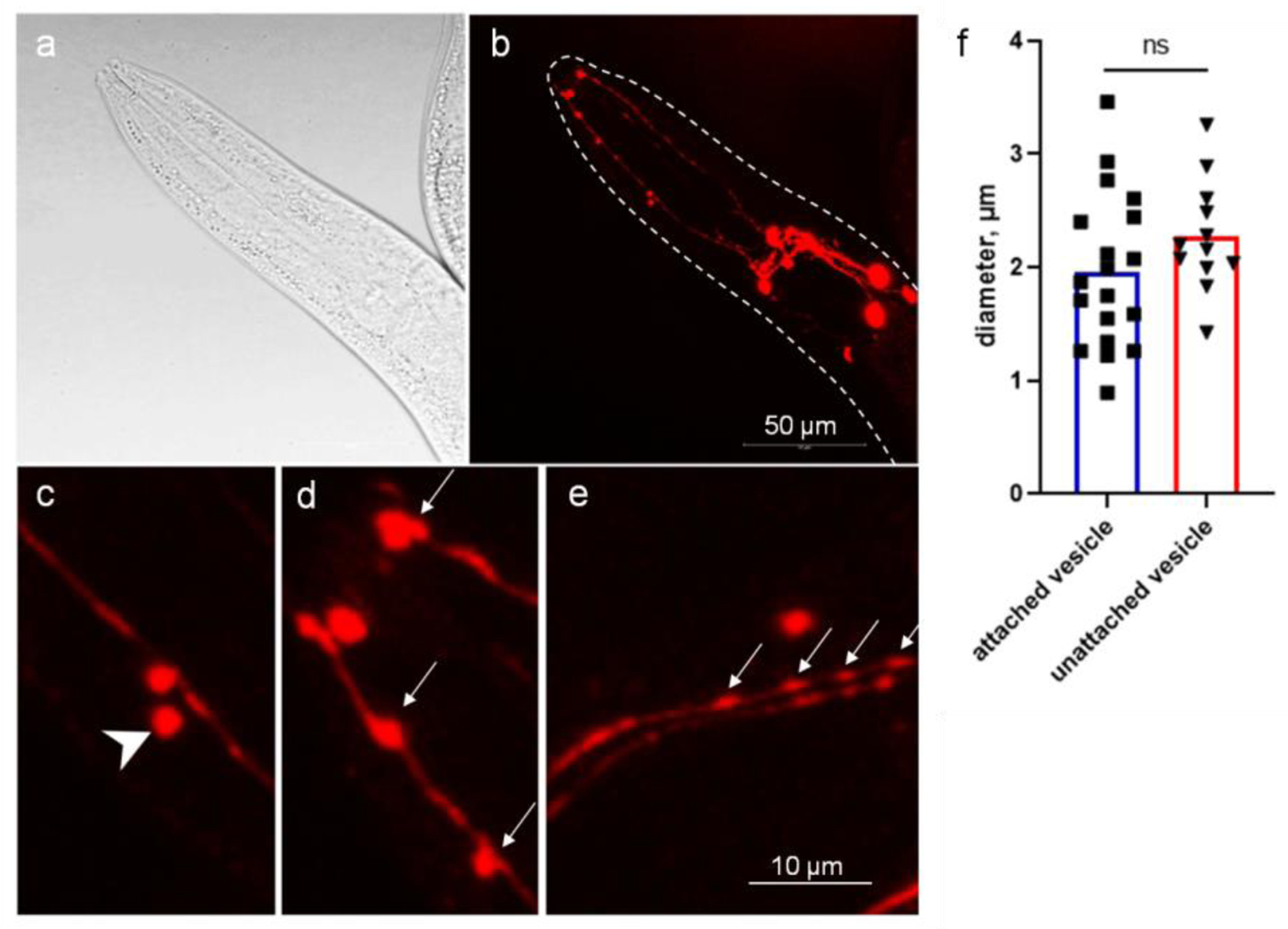
Figure 1.

C. elegans CEP dendrites produce microvesicles. Images of adult stage of the OH7193 strain were acquired by Leica SP8 confocal microscope. a, bright field image of the head region. b, corresponding image of DAergic neurons in the head region. c, unattached vesicle shed from the CEP dendrite. d and e, attached vesicle on the CEP dendrites. Arrow head shows unattached vesicle; arrows show attached vesicle. f, diameters of the vesicular structures in the CEP dendrites. t-test.
