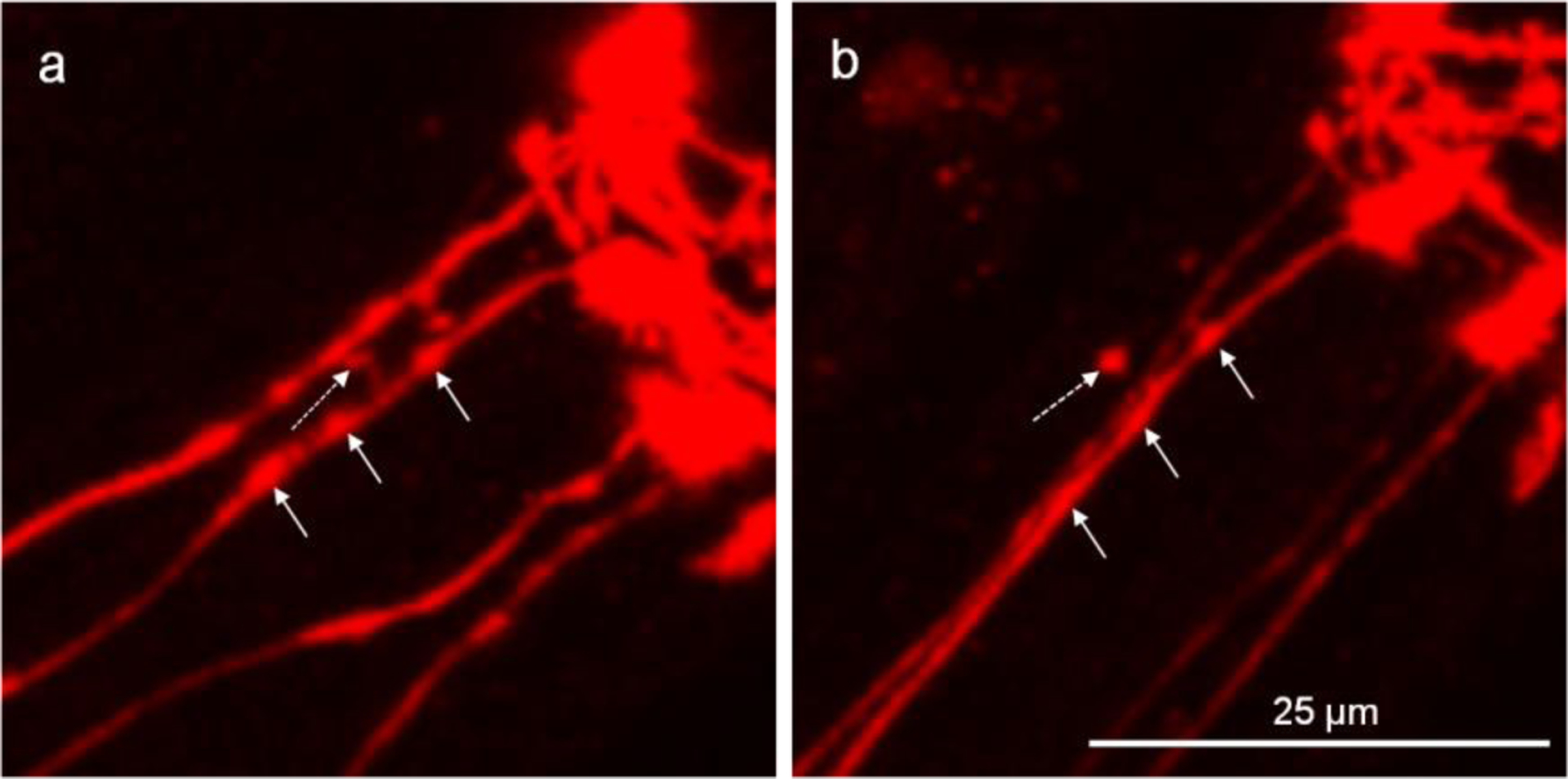
Figure 2.

Attached vesicles shed off the CEP dendrite to become unattached vesicles. The head region of DAergic neurons was imaged to show the four parallel CEP dendrites. a, the dotted-line arrow shows the attached vesicle which is linked to the dendrite with a thin tubular structure, and the other straight-line arrows show the areas that are likely to form attached vesicles. b, of the same worm from “a” after a 4-h period, shows the attached vesicle shed off from the dendrite, while the areas that are likely to form attached vesicles become flattened.
