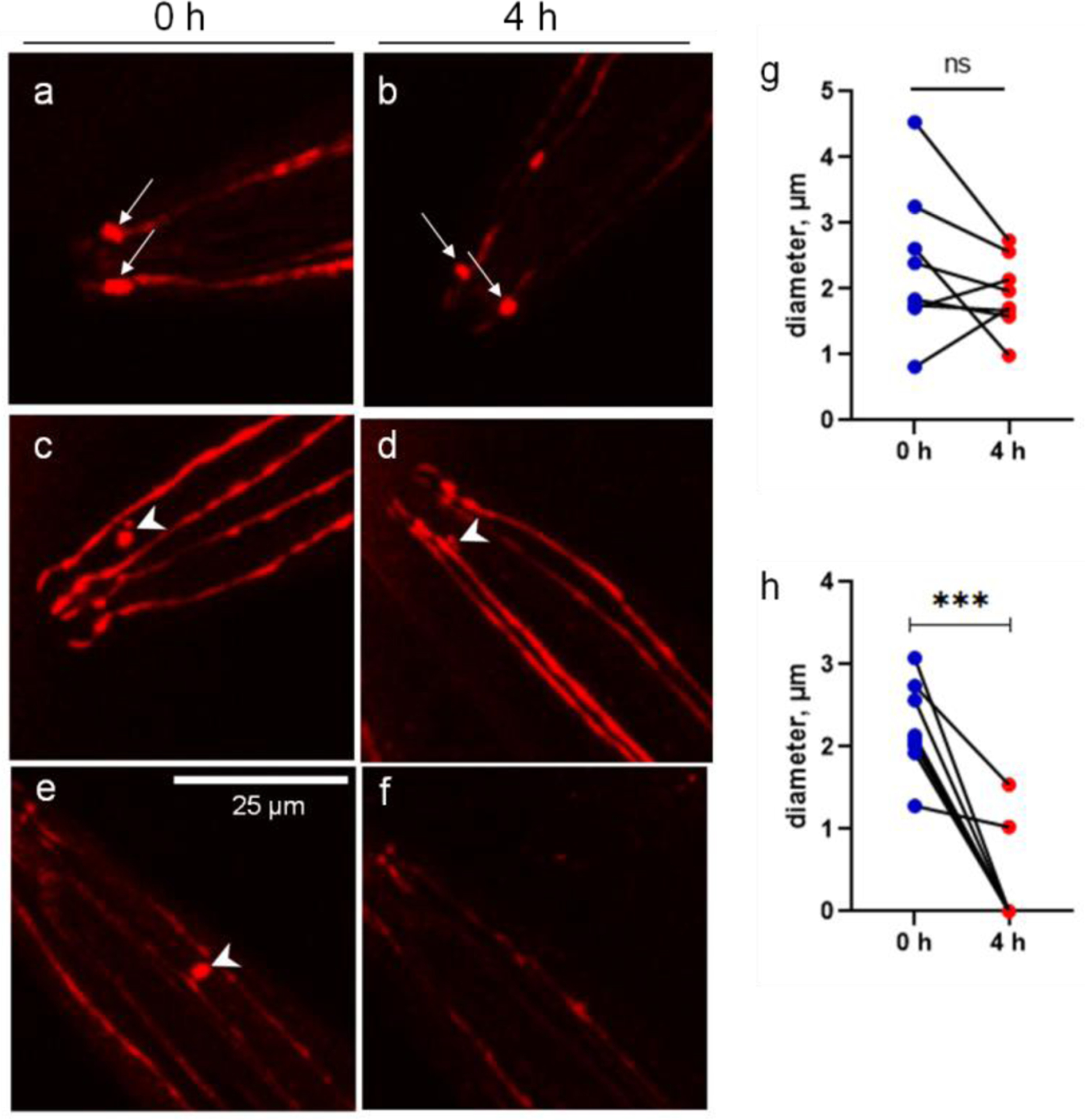
Figure 3.

The CEP vesicles are dissolved during a 4-h period. The images of single-worm tracking show the dynamics of attached vesicles (arrow) and unattached vesicles (arrow head). a and b, show two attached vesicles at the CEP ciliary bases in the proximity of mouth area. c and d, show the unattached vesicle becomes much smaller after a 4-h period. e and f, show the unattached vesicle becomes invisible after a 4-h period. g, diameters of attached vesicles at 0 and 4 h. h, diameters of unattached vesicles at 0 and 4 h. ***p<0.001, paired t-test.
