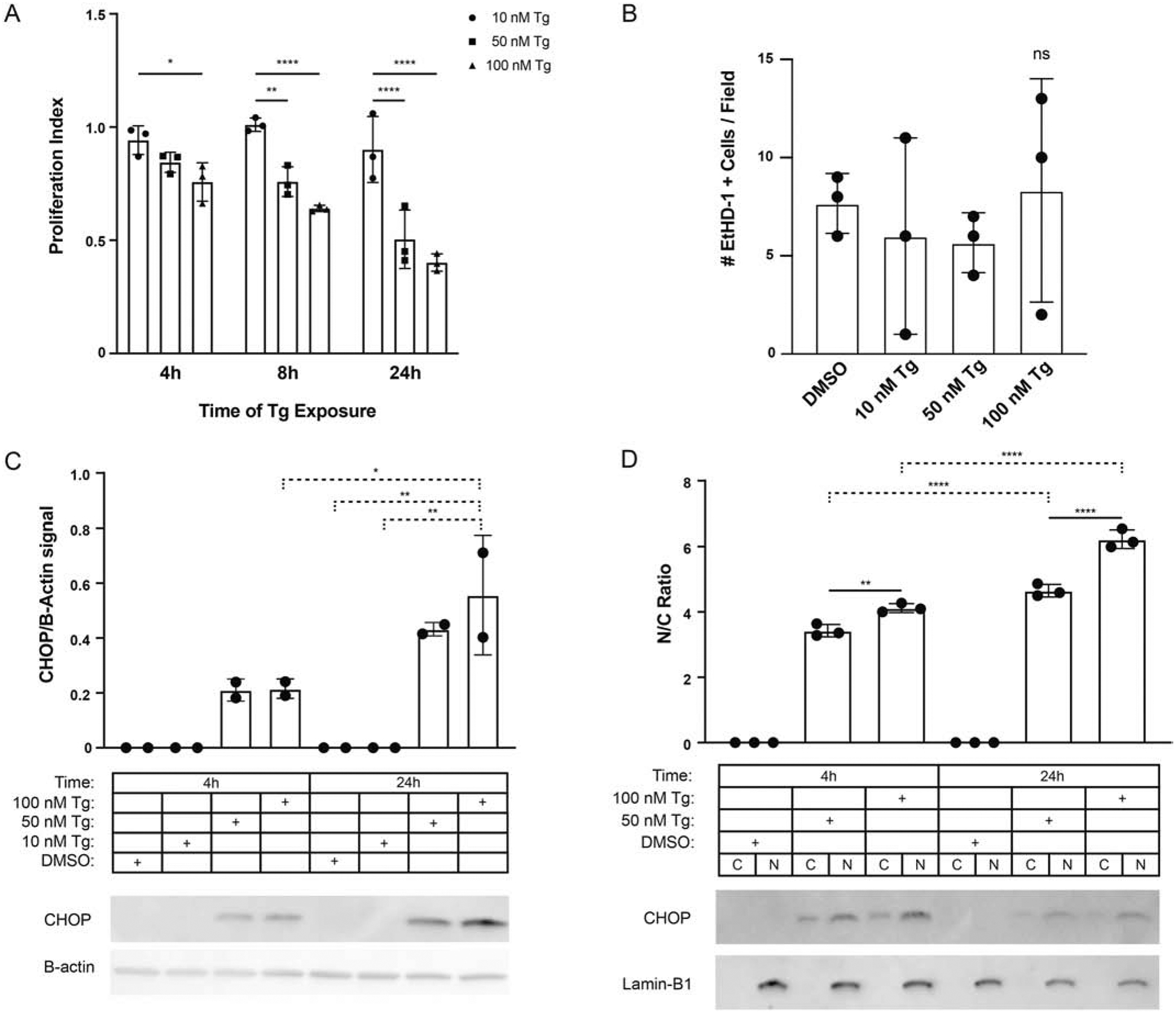
Fig. 1. Thapsigargin induces CHOP in both the nuclear and cytoplasmic compartments.

(A) Proliferation analyses of Vero cultures at 4, 8, and 24h exposed to thapsigargin (Tg) 10, 50, and 100nM. The proliferative index represents the number of nuclei in Tg treated wells normalized to DMSO; n=3. (B) Effects of Tg treatment on cell injury measured as the total number of ethidium homodimer (EthD-1) positive cells per field; n=3. (C) Western analyses for CHOP and β-actin expression in whole-cell lysates following exposure to 10, 50, or 100 nM Tg vs. DMSO control at 4 and 24 hours. Values reflect CHOP signal by densitometry normalized to β-actin; n=2. (D) Western analyses for CHOP and Lamin-B1 expression nuclear and cytoplasmic cell fractions from Vero cultures treated with 50 and 100nM thapsigargin or DMSO control at 4 and 24 hours. Values reflect the ratio of nuclear to cytoplasmic CHOP signal by densitometry; n=3. * p ≤ 0.05, ** p ≤ 0.01, **** p ≤ 0.0001.
