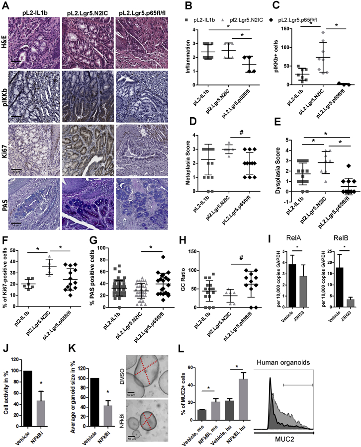
Figure 7.

A RelA knockout reduces the pathology observed in mice with induced inflammation and Notch signaling. A) Representative IHC images of RelA KO mice that are depicted next to L2-IL1B and pL2.Lgr5.N2IC mice. (B-H) Histopathological evaluations for metaplasia, dysplasia, inflammation, GC ratio, and numbers of pIKK+, Ki67+ and PAS+ cells in the BE region in 10 high-power fields. Statistical analysis was performed using one-way ANOVA and Tukeys multiple comparison test, *p<.05, or the Kruskal-Wallis test #p<.05. All indicated IHC based results were derived from mice ranging from 10 to 12 months of age. Only mice with the same age were compared. (I-L) L2-IL1B mice derived organoid cultures were treated with the NF-κB inhibitor, JSH-23 (10μM), or vehicle control. (I) qRT-PCR of L2-IL1B mice derived organoids. (J) Cell activity was measured via MTT assay and (K) organoid growth was determined microscopically. (L) Flow cytometry of organoid cells after enzymatic disaggregation to determine the proportion of Muc2+ cells, derived from (ms; L2-IL1B) and human (hu, BE) samples. The representative histogram indicates the range of MUC/Muc2+ cells for vehicle (dark, 22.5%) and inhibitor (light grey, 41.1%) treated conditions. Data is presented as means ± standard deviation of at least three independent experiments. Statistical analysis was performed using Student's t-test, *p<.05
