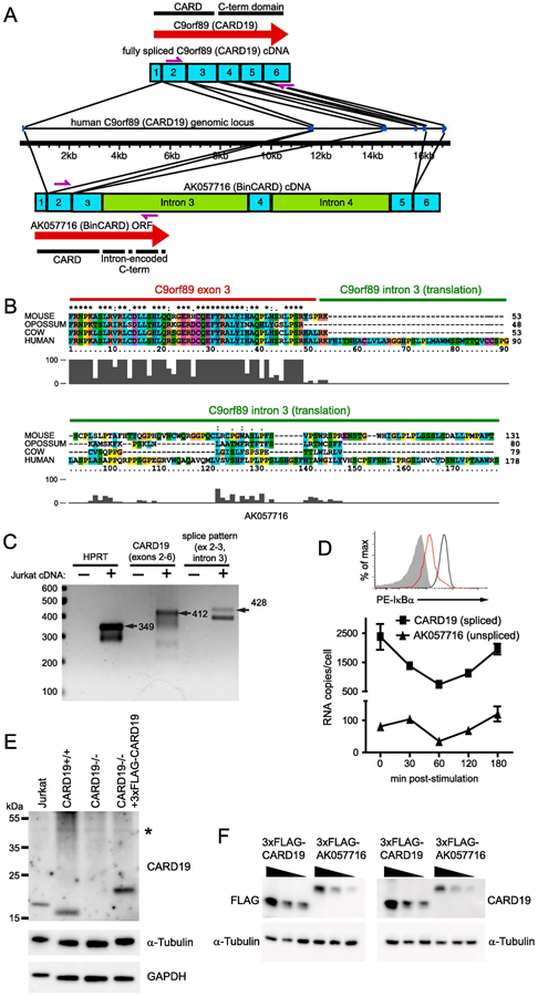
Figure 2. The AK057716 cDNA clone is the product of an incompletely spliced CARD19 mRNA.

A, Diagram comparing the structures of the fully spliced CARD19 transcript and the partially spliced AK057716 transcript. The human CARD19 (c9orf89) gene structure is indicated in the middle of the panel, with transcript structures shown above and below the gene. Exons are designated by numbered blue boxes and unspliced introns are green. Locations of ORFs are designated by red arrows and primers used for PCR in C are indicated by purple arrows. B, Translated sequences from the intron 3/exon 3 boundary region of the human CARD19 gene and homologues from mouse, opossum, and cow. Clustal-X2 alignment shows an intronic translation of variable length with almost no homology beyond the splice junction. C, cDNA from the Jurkat T cell line was amplified by PCR to detect HPRT, exons 2-6 (fully spliced) of CARD19, and the exon 2-exon 3-intron 3 splicing pattern of the AK057716 clone (see A for locations of primers for CARD19 and AK057716). Expected products of the indicated lengths were detected with each of the primer sets. D, Jurkat T cells were stimulated with anti-CD3 plus anti-CD28 and RNA was harvested at the indicated times post-stimulation. Cells from the 30 min time point were analyzed by intracellular flow cytometry to confirm efficient degradation of IκBα at 30 min post-stimulation, indicative of efficient activation of the TCR (top). Real-time PCR analysis was performed on each sample using primers to specifically detect the CARD19 splicing pattern (exon 3-exon 4) and the AK057716 splicing pattern (exon 2-exon 3-intron 3), and signals were compared to plasmid standard curves to determine per cell copy number at each post-stimulation time point (bottom). E, anti-CARD19 was used in an immunoblotting experiment to detect endogenous CARD19, α-tubulin, and GAPDH in Jurkat T-cells, murine CARD19+/+ and CARD19−/− CD8 T cells, and ectopic 3×FLAG-CARD19 in retrovirally-transduced CARD19−/− CD8 T cells. F, HEK293T cells were transfected with decreasing concentrations of 3xFLAG-CARD19 or 3×FLAG-AK057716 plasmid. Lysates were separated via SDS-PAGE and probed with either anti-FLAG (left) or anti-CARD19 (right). Both sets of lysates were probed with anti-α-tubulin.
