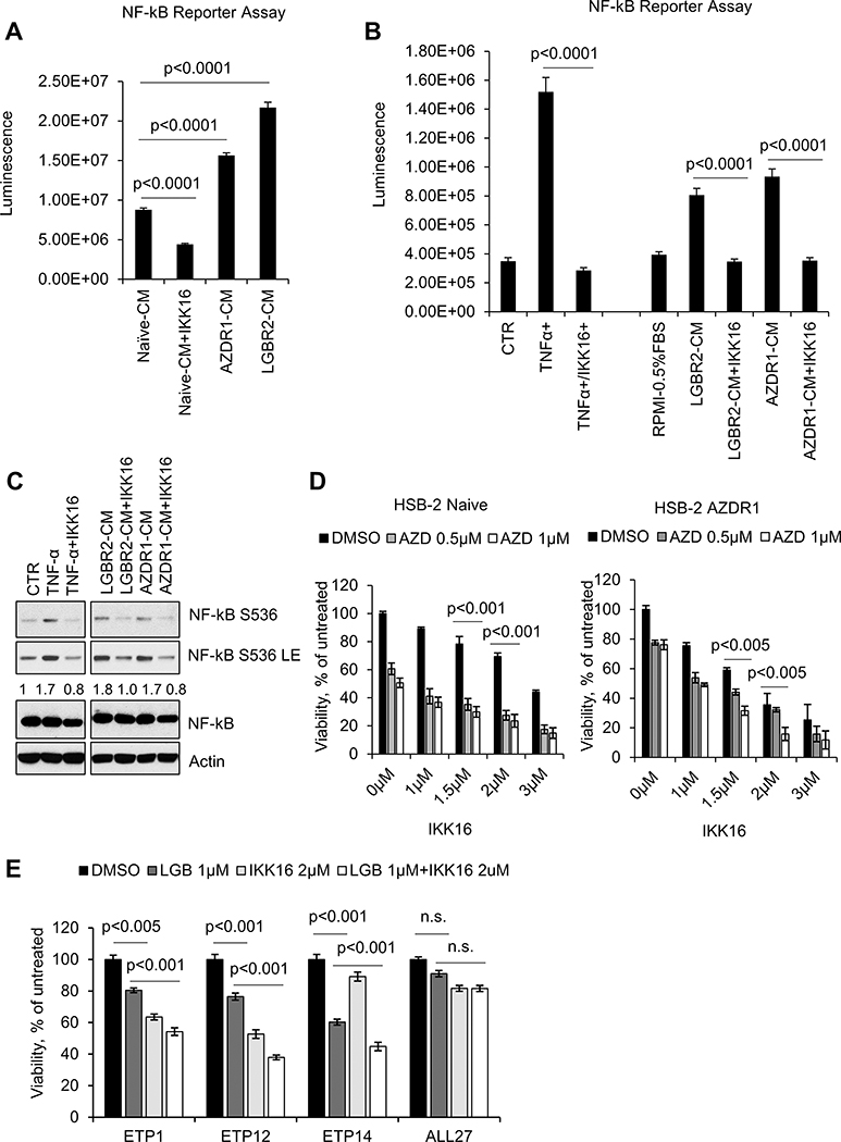
Figure 6. NF-κB activation in PIMi treated HSB-2 cells.
(A) HEK-293T-NF-κB-luc cells were incubated with conditioned media from HSB-2 naïve or AZDR1 and LGBR2 cells with or without IKK16 (2 μM) for 6h. (B & C) HEK-293T-NF-κB-luc cells were incubated with media containing TNF-α (20 ng/mL) alone or in combination with IKK16 (2 μM) or CM from HSB-2 LGBR2 or AZDR1 with or without IKK16 (2 μM) for 6h. NF-κB mediated luciferase activity was measured using One-Glo Luciferase Assay System. Cell lysates were Western blotted with the specified antibodies. LE: long exposure. (D) Cell viability of HSB-2 naïve and AZDR1 cells treated with indicated concentrations of AZD alone and/or in combination with IKK16 for 72h. (E) T-ALL PDX cells were incubated with the indicated concentrations of LGB321 (LGB) alone or in combination with IKK16 for 72h and then the percentage of viable cells was quantified by the ATPlite assay. For (D and E), the growth of DMSO control cells was considered 100% and percent cell growth after individual treatment is reported relative to the DMSO. The data shown are the average +/− S.D. of three independent experiments.

