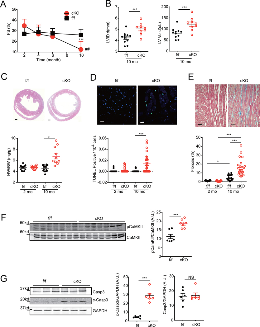
Figure 1. Cardiac deletion of SAP97 promotes spontaneous heart failure in ageing mice.
A) Cardiac fraction shortening from SAP97-f/f and SAP97-cKO mice were measured with echocardiogram at different ages. Data were from 11 SAP97-f/f and 9 SAP97-cKO mice. ## p < 0.01 by two-way ANOVA followed by Tukey’s test. B) Cardiac LVID and LV volume on 10-month old mice are plotted. ***p < 0.001 between SAP97-cKO and SAP97-f/f mice at 10-month age. C-E) Representative images show H & E staining (Scale bar 500 μm), TUNEL staining (Scale bar 10 μm), Masson’s trichrome staining (Scale bar 100 μm) of heart tissues from SAP97-cKO and SAP97-f/f at 10-month old. Nuclei are shown with DAPI staining. The quantification of the images are plotted below. For H &E staining, * p < 0.05 by one-way ANOVA followed by Tukey’s test. For TUNEL staining, data were 2–3 repeated measurements from 5 2-month and 6 10-month old mice; and Masson’s trichrome staining, data were 2–3 repeated measurements of 5 2-month and 7 10-month old mice. * p < 0.05 and *** p < 0.001 by one-way nested ANOVA followed by Tukey’s test. F-G) Western blots show total and phosphorylated CaMKII and total and cleaved caspase 3 in 10-month old SAP97-cKO and SAP97-f/f heart tissues. ***p < 0.001 by unpaired student t-test.

