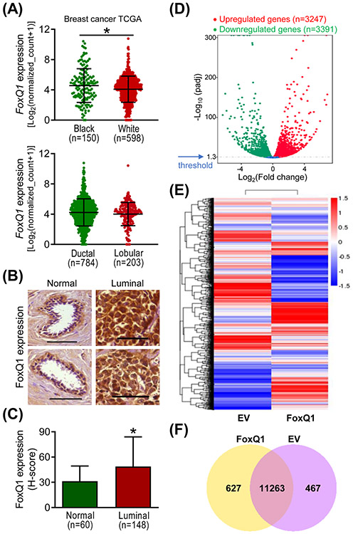
Figure 1.
FoxQ1 expression was significantly higher in luminal-type human breast cancer specimens compared to normal human mammary tissues. A, Expression of FoxQ1 in mammary tumors of black and white women, and in ductal and lobular carcinomas. B, Representative immunohistochemical images (200× magnification; scale bar = 100 μm) for FoxQ1 expression in normal human mammary tissues and luminal-type human breast cancer tissues. C, Quantification of FoxQ1 expression in normal human mammary tissues and luminal-type human breast cancer tissues. Results shown are mean ± SD. *P < 0.05 by two-sided Student's t-test. D, Volcano plot for differential gene expression. E, Heatmap of the differentially expressed genes in empty vector transfected cells (EV) and FoxQ1 overexpressing SUM159 cells. F, Venn diagram showing unique and overlapping genes between EV and FoxQ1 overexpressing SUM159 cells.

