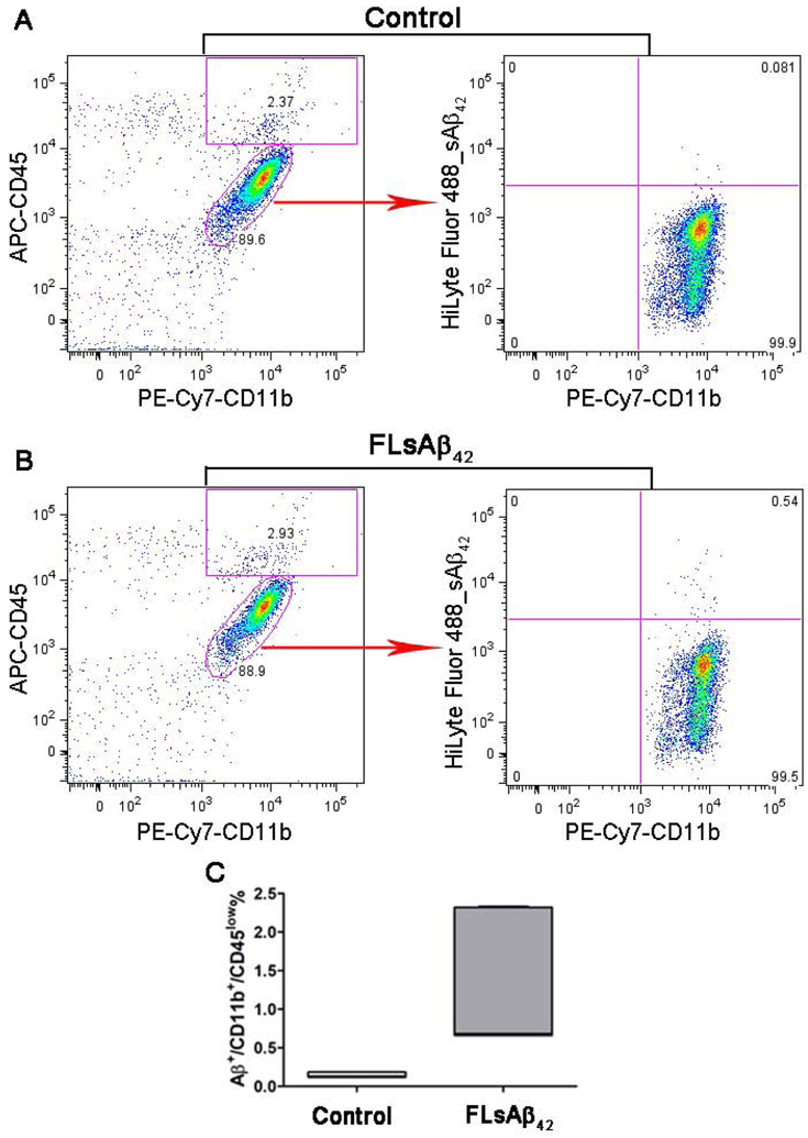
Figure 3. The uptake of microinjected synthetic sAβ by microglia was measured by flow cytometry.
(A-C) Five days after surgery, adult primary microglia were isolated and stained with PE-Cy7-CD11b and APC-CD45. The CD11b+/CD45low microglia were gated and the percentage of Aβ+ cells among those gated microglia was analyzed. Representative Aβ+/CD11b+/CD45low microglia were recorded in the control group microinjected with fluorescence-labeled scrambled peptide (A) and in the treatment group microinjected with FLsAβ42 (B). (C) The percentage of Aβ+/CD11b+/CD45low microglia in both groups was analyzed (n= 5 females per group); p < 0.05 (nonparametric Mann Whitney test). Detailed statistical results are shown in Table 2. Only a very small fraction (less than 1.5%) of microglia in the FLsAβ42-treated group were Aβ-positive.

