
Figure 5. Natural sources of sAβ cannot be taken up by N9 microglial cells but monomer degradation can be reversed by an insulin degrading enzyme inhibitor.
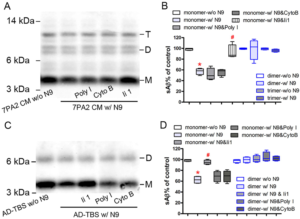
(A) and (C) N9 microglial cells were pretreated with or without 50 μg/ml polyinosinic acid (Poly I), 20 μg/ml cytochalasin B (Cyto B), or 10 μM Ii 1 and then incubated with or without soluble Aβ (sAβ from 7PA2 CM (A) or AD-TBS brain homogenate (C) for 3 h. Conditioned medium were collected, pre-cleared with protein A-Sepharose, and immunoprecipitated overnight with anti-Aβ antiserum (R1282). Aβ was detected in the immunoprecipitate by an anti-Aβ antibody (6E10) using Western blot assay. (B) and (D) The percentage of different forms of sAβ in panel A and C was quantified, respectively. The data, expressed as percentages of control (i.e. w/o N9), represent the means ± SEM of four separate experiments from each A and C; * p < 0.05 versus control, # p < 0.05 versus the group w/ N9 alone (nonparametric Mann Whitney test). Detailed statistical results are shown in Table 2.