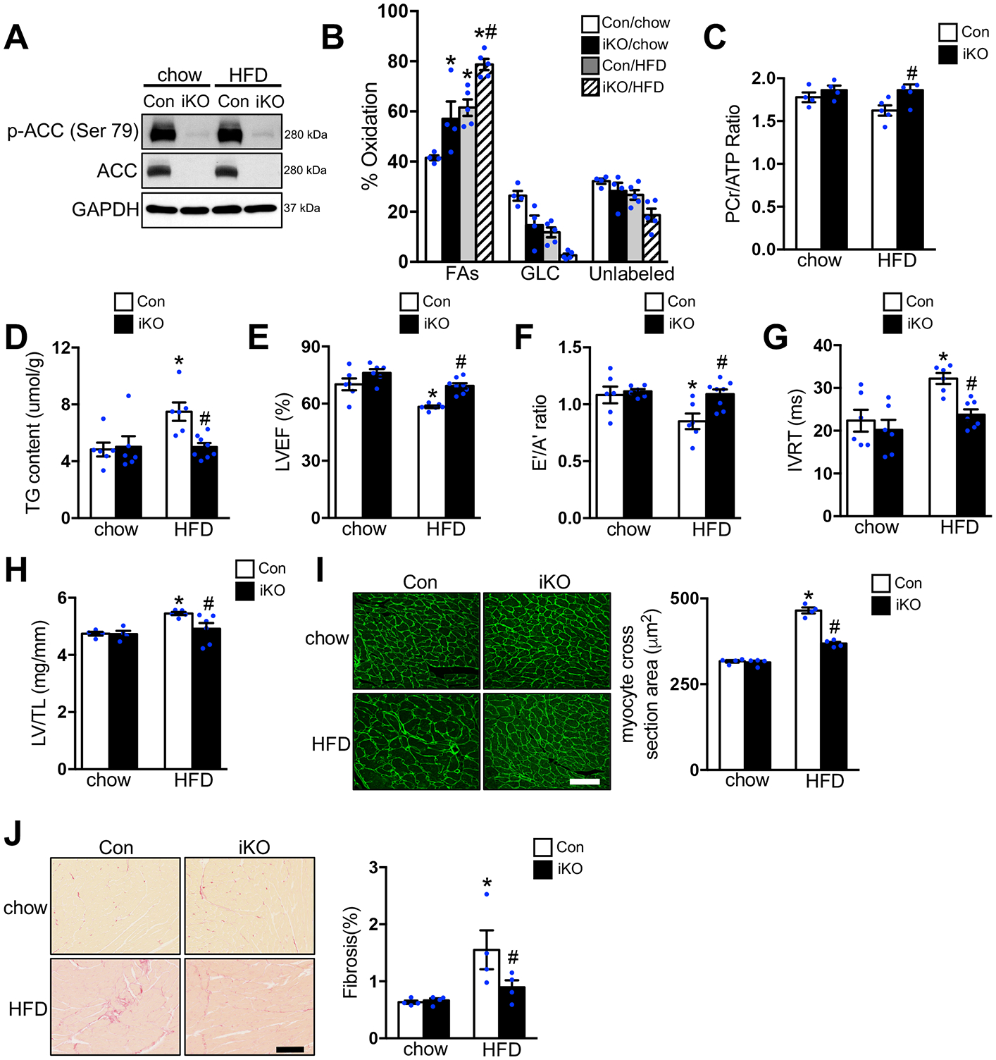
Figure 1. Increasing FAO attenuated HFD induced cardiomyopathy.

Con and ACC2 iKO mice were subjected to chow or HFD feeding for 24 weeks. (A) Representative immunoblots of heart lysates of p-ACC (Ser 79), ACC and GAPDH are shown. (B-C) Hearts were perfused with a buffer containing 13C labeled fatty acids, 13C labeled glucose, lactate, and insulin (mixed substrate). (B) Contribution of 13C labeled substrates to tricarboxylic acid (TCA) cycle was determined by 13C NMR spectroscopy in heart extracts. Relative contribution of fatty acids (FAs), glucose (GLC), and other unlabeled substrates (lactate, endogenous) (Unlabeled) is shown (*p<0.05 vs. Con/chow, #p<0.05 vs. Con/HFD, n=4–5). (C) Phosphocreatine to ATP (PCr/ATP) ratio was measured by 31P NMR spectroscopy in isolated perfused hearts (#p<0.05 vs. Con/HFD, n=4–5). (D) Cardiac triglyceride (TAG) content normalized to tissue weight (*p<0.05 vs. Con/chow, #p<0.05 vs. Con/HFD, n=6–8). (E-G) Left ventricular ejection fraction (LVEF), E’/A’ ratio and isovolumic relaxation time (IVRT) were measured by echocardiography (*p<0.05 vs. Con/chow, #p<0.05 vs. Con/HFD, n=6–8). (H) The left ventricle weight to tibia length (LV/TL) ratio of ACC2 iKO and Con mice under chow- or HFD-fed conditions (*p<0.05 vs. Con/chow, #p<0.05 vs. Con/HFD, n=4–6). (I) Representative wheat germ agglutinin (WGA) staining and quantification of cardiomyocyte cross-sectional area in indicated hearts after 24 weeks’ chow or HFD feeding (*p<0.05 vs. Con/chow, #p<0.05 vs. Con/HFD, n=4), Scale bar, 50μm. (J) Representative picric acid sirius red (PASR) staining and quantification of the percentage of fibrosis in indicated hearts after 24 weeks’ chow or HFD feeding (*p<0.05 vs. Con/chow, #p<0.05 vs. Con/HFD, n=4), Scale bar, 100μm.
