
FIGURE 1.
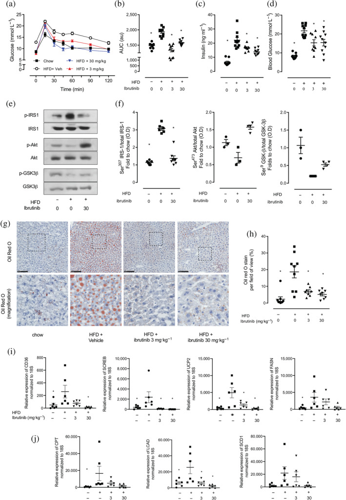
Ibrutinib treatment restored insulin signalling through IRS‐1/Akt/GSK‐3β in mice fed a HFD. (a) Oral glucose tolerance (OGTT) was assessed over 120 min, 1 week prior to harvest. (b) The AUC of OGTT was calculated for respective groups and used for statistical analysis. (c) Plasma insulin levels were measured in plasma isolated from whole blood at harvest. (d) Basal, non‐fasted, blood glucose was measured at week 11 1 h prior to harvest. Data shown are (in a) means ± SEM in b, c, d, with individual values; n = 9‐10 per group. * P < 0.05, significantly different from HFD + Veh; one‐way ANOVA with Bonferroni post hoc test. (e) Representative western blots for phosphorylation of Ser307 on IRS‐1 in the liver and normalized to total IRS‐1; for phosphorylation of Ser473 on Akt in the liver and normalized to total Akt; for phosphorylation of Ser9 on GSK‐3β in the liver and normalized to total GSK‐3β and (f) quantified using densitometry. Data shown are individual values with means ± SEM; n = 3–6 per group. * P < 0.05, significantly different from HFD + Veh; one‐way ANOVA with Bonferroni post hoc test. (g) Representative images of hepatic lipid deposition assessed by Oil Red‐O staining and (h) quantified. Scale bars measure 50 μm. Inset images are 4× digital zoom. (i) Relative gene expression of CD36, SCREB, UCP2, and FASN were assessed by qPCR and normalized to 18S. (j) Relative gene expression of CTP, LCAD, and SCD1 were assessed by qPCR and normalized to 18S. and gene expression data. Data shown are individual values with means ± SEM; n = 6 per group. * P < 0.05, significantly different from HFD + Veh; one‐way ANOVA with Bonferroni post hoc test