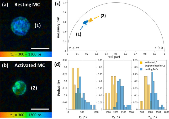
Figure 2.
TPE-FLIM parameters of hsMCs in vitro. TPE-FLIM τm image (mean fluorescence lifetime τm in the 300–1,300 ps range) of hsMCs in vitro: Primary human skin MCs were prepared from skin obtained from breast reduction surgery and cultured for 5 days. (a) MCs were washed with PBS and imaged directly or (b) were incubated with ionomycin at 1 µM for 15 min before imaging. TPE-FLIM parameters τ1, τ2 and τm were recorded with laser excitation at 760 nm with 100 fs pulses and a repetition rate of 80 MHz at 3–5 mW. Scale bar: 10 µm. (c) Phasor plot showing distribution of τm for resting (blue, MC (1)) and ionomycin-treated, activated (orange, MC (2)) hsMCs. (d) Distribution of TPE-FLIM parameters τ1, τ2 and τm for resting (blue, n = 43) and ionomycin-treated, activated (orange, n = 13) MCs.

