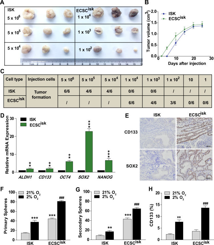
Fig. 3. Characterisation of the increased stemness properties in ECSCs in vivo.
a, b Tumorigenicity detected by the morphology (a) and volumes of xenografts (b) formed from ISK cells and ECSCisk. c Tumorigenicity initiating capacity of the minimum number of ISK cells and ECSCisk. d qRT-PCR analysis of the expression of ALDH1, CD133, OCT4, SOX2 and NANOG in the tumours of the two groups. e Immunostaining of CD133 and SOX2 in tumours by IHC. f, g The numbers of primary (f) and secondary (g) spheres formed from ISK cells and ECSCisk under hypoxic or normoxic conditions for 72 h. h FACS analysis of the percentages of CD133-positive cells in tumours from ISK cells and ECSCisk in hypoxic or normoxic conditions for 72 h. **P < 0.01, and ***/###P < 0.001.

