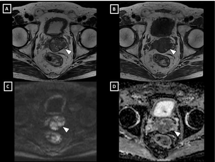
Figure 1.
MRI findings of the prostate tumour. T2-weighted (A) and T1-weighted (B) images revealed the tumour, which showed expansive progression with a clear capsule. The tumour presented with high signal intensity on diffusion-weighted images (C) and had low apparent diffusion coefficient values (D).

