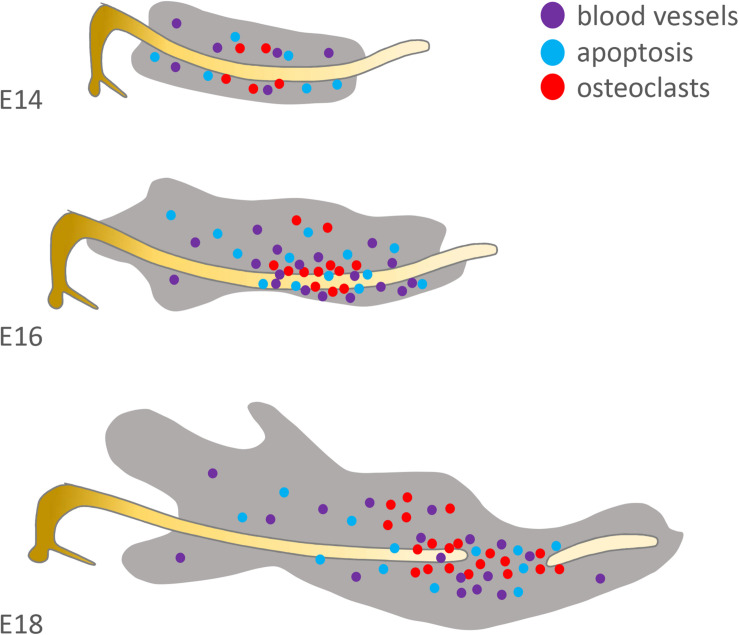
FIGURE 4.
Distribution of factors suspected to be involved in Meckel’s cartilage degradation. Original schematic showing localisation of osteoclasts/osteoclastic precursors (red = TRAP+), endothelial cells (purple = CD31+) and apoptotic cells (blue = TUNEL+) in the mouse at E14, 16 and 18. Based on the published literature (see Harada and Ishizeki, 1998; Sakakura et al., 2005; Yang et al., 2012).

