. 2020 Sep 10;2(5):e200276. doi: 10.1148/ryct.2020200276

Figure 3.

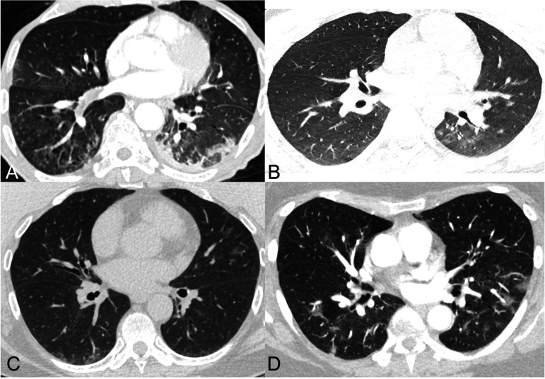
Examples of cases for which there was significant disagreement in assignment of RSNA consensus COVID-19 category.A) 67-year-old man with clinical signs of pneumonia and 4 negative PCRs for COVID-19 with sputum samples positive for streptococcus pneumoniae. Axial CT image shows a combination of tree-in-bud centrilobular nodules in the lower lobes and peripheral ground glass opacity and consolidation in the left lower lobe. Categories 3, 2, and 1 were assigned by 4, 3, and 2 readers respectively.B) year-old man with 2 negative PCR results for SARS-CoV-2 and presumed aspiration or non-23-COVID-19 infection. Axial CT image shows minimal patchy ground glass opacities in the left lower lobe; there was a question of atelectasis or subtle peripheral ground glass opacity in the posterior right lower lobe. Categories 3, 2, 1, and 0 were assigned by 1, 6, 1, and 1 readers respectively.C) 64-year-old woman with PCR-proven COVID-19 pneumonia who presented with fever, productive cough, fatigue, and anosmia. Axial CT image shows patchy ground glass opacities in the lingula and a small amount of peripheral ground glass opacity and atelectasis in the posterior lower lobes. Categories 3, 2, 1, and 0 were assigned by 4, 3, 1, and 1 readers respectively. Reasons given by readers for uncertainty included doubts about peripheral distribution, and difficulty in classification in the setting of minimal disease and posterior atelectasis.D) 65-year-old woman with PCR-proven COVID-19 pneumonia who presented with palpitations, back pain, and low-grade fevers. Axial CT image shows patchy ground glass opacities bilaterally. Categories 3 and 2 were assigned by 5 and 4 readers respectively. Reasons given by readers for uncertainty included difficulty in classifying as peripheral or diffuse and questionable morphology of the ground glass opacities.

Examples of cases for which there was significant disagreement in assignment of RSNA consensus COVID-19 category.

A) 67-year-old man with clinical signs of pneumonia and 4 negative PCRs for COVID-19 with sputum samples positive for streptococcus pneumoniae. Axial CT image shows a combination of tree-in-bud centrilobular nodules in the lower lobes and peripheral ground glass opacity and consolidation in the left lower lobe. Categories 3, 2, and 1 were assigned by 4, 3, and 2 readers respectively.

B) 23-year-old man with 2 negative PCR results for SARS-CoV-2 and presumed aspiration or non-23-COVID-19 infection. Axial CT image shows minimal patchy ground glass opacities in the left lower lobe; there was a question of atelectasis or subtle peripheral ground glass opacity in the posterior right lower lobe. Categories 3, 2, 1, and 0 were assigned by 1, 6, 1, and 1 readers respectively.

C) 64-year-old woman with PCR-proven COVID-19 pneumonia who presented with fever, productive cough, fatigue, and anosmia. Axial CT image shows patchy ground glass opacities in the lingula and a small amount of peripheral ground glass opacity and atelectasis in the posterior lower lobes. Categories 3, 2, 1, and 0 were assigned by 4, 3, 1, and 1 readers respectively. Reasons given by readers for uncertainty included doubts about peripheral distribution, and difficulty in classification in the setting of minimal disease and posterior atelectasis.

D) 65-year-old woman with PCR-proven COVID-19 pneumonia who presented with palpitations, back pain, and low-grade fevers. Axial CT image shows patchy ground glass opacities bilaterally. Categories 3 and 2 were assigned by 5 and 4 readers respectively. Reasons given by readers for uncertainty included difficulty in classifying as peripheral or diffuse and questionable morphology of the ground glass opacities.