Figure 1.

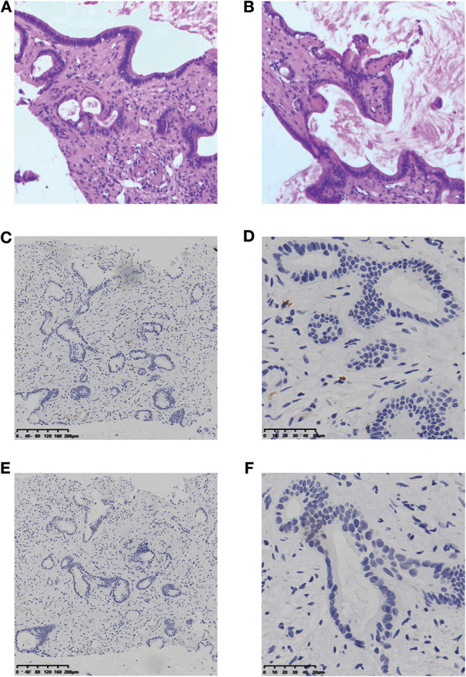
H&E staining (A, B) and IHC result of CD8 (C,D) and PD-L1 (E,F) for the liver lesion. H&E staining, hematoxylin and eosin staining; IHC, immunohistochemistry; PD-L1, programmed cell death ligand 1.

H&E staining (A, B) and IHC result of CD8 (C,D) and PD-L1 (E,F) for the liver lesion. H&E staining, hematoxylin and eosin staining; IHC, immunohistochemistry; PD-L1, programmed cell death ligand 1.