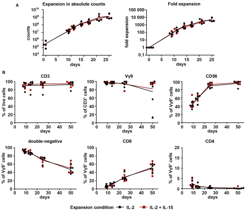
Figure 1.
Proliferation and purity analysis of Vγ9Vδ2 T cells expanded with IL-2 (1,000 U/ml) or combined IL-2 (100 U/ml) + IL-15 (100 U/ml). Vγ9Vδ2 T cells were expanded with 10 μM ZOL and either (a) 1000 U/ml IL-2 (black) or (b) 100 U/ml IL-2 + 100 U/ml IL-15 (red). (A) Total cell count of Vγ9Vδ2 T cell cultures expanded from PBMCs (n = 4, compiled from three independent experiments; γδHD1–4) for 25 days. Fold expansion was calculated based on the number of Vγ9+ cells initially present in the culture. (B) Vγ9Vδ2 T cell cultures expanded from PBMCs (n = 6, compiled from three independent experiments; γδHD5–10) for 50 days were analyzed by flow cytometry for expression of CD3, Vγ9, CD56, CD4, and CD8 on days 9, 14, 25, and 50 of culture. Lymphocytes were gated first, and then doublets were excluded before gating on live cells. Lines represent means of all cultures.

