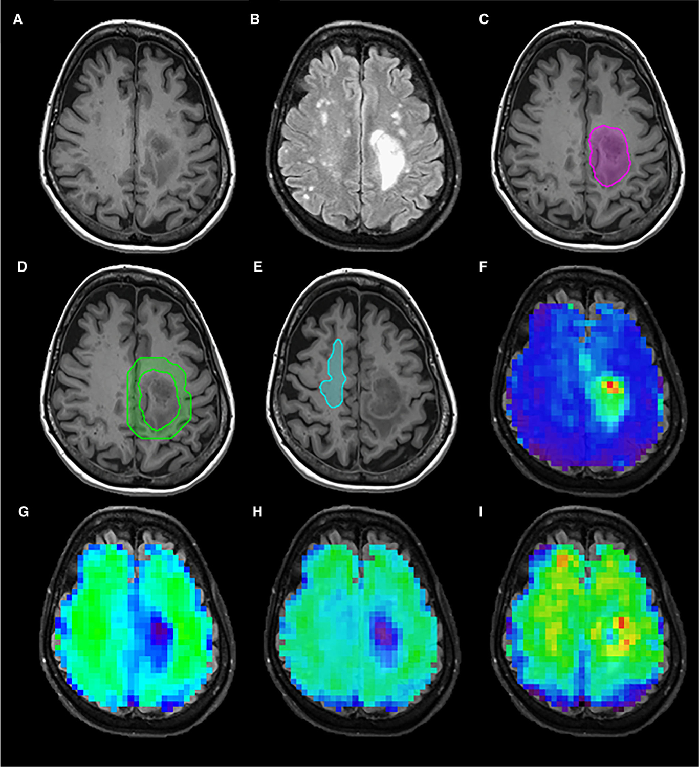
Fig 1.

T1 postcontrast image (A), Fluid-attenuated inversion recovery image (B), volume of interests for active tumor (C), peritumoral region (D), contralateral normal-appearing white matter (E), and MRSI metabolite maps for Cho/NAA (F), NAA (G), Creatine (H), and Choline (I) in a 70-year-old female IDH wild-type subject.
