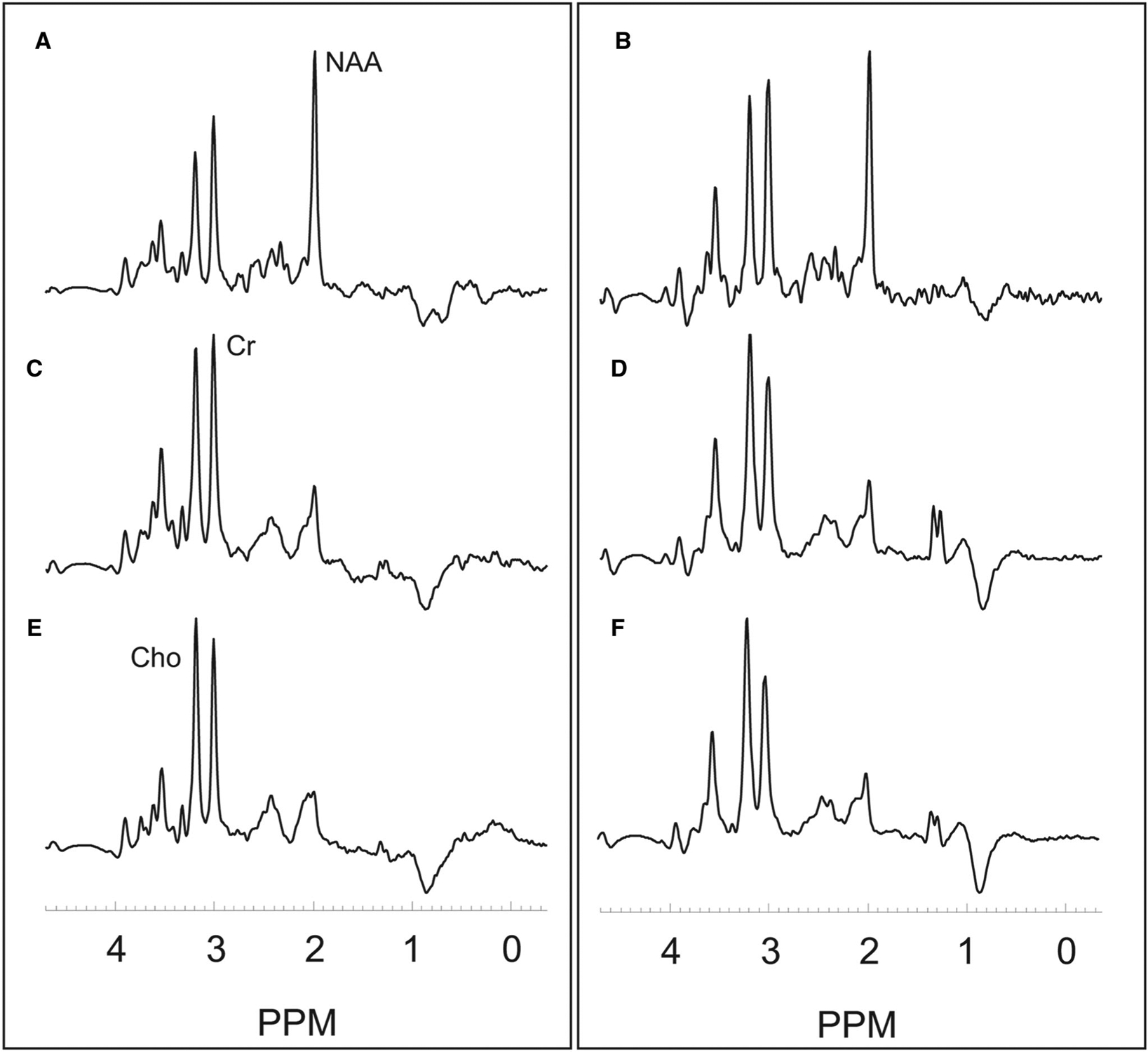
Fig 2.

Example integrated spectra for the volume of interests for contralateral normal-appearing white matter (A,B), peritumoral region (C,D), and active tumor (E,F) in a 70-year-old female with an IDH wild-type Grade 4 glioma with imaging data shown in Figure 1 (left panel) and in a 53-year-old female with an IDH mutant Grade 3 oligodendroglioma (right panel). ppm denotes parts per millions with NAA at 2.0 ppm, Cho at 3.2 ppm, and Cr at 3.0 ppm.
