Figure 3.
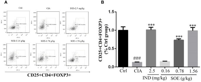
SOE upregulated the level of CD25+CD4+FOXP3+ cells in spleen. The purified splenocytes (5–10 × 105 cells) were surface-stained with anti-rat CD4-FITC/CD25-PE for 30 min on ice in darkness, then permeabilized and stained with anti-mouse/rat/human Foxp3-Alexa Fluor® 647 for another 30 min. The acquisition of flow cytometry data and the analysis of a cell population of at least 1 × 10⁴ splenocytes were performed on flow cytometer with the BD FACSDiva Software (FACSCanto, BD Biosciences). (A) Representative images of flow cytometry for CD4+CD25+Foxp3+ regulatory T cells. (B) Quantification of the number of CD4+CD25+Foxp3+ regulatory T cells. (###P < 0.001 vs. Ctrl group; ***P < 0.001 vs. CIA group, n = 6).

