
Fig 2. 3D IVM set up and cumulus expansion assessment.
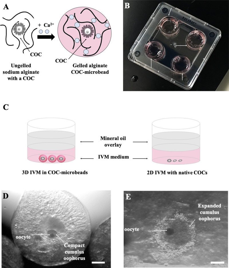
(A) Calcium-induced alginate gelification in presence of a COC to produce a COC-microbead. (B) COC-microbeads placed in 4-well culture dishes for IVM (white arrows). (C) Diagram of oocytes cultured under 3D and 2D IVM conditions. (D, E) Representative images showing a 1% alginate COC-microbead as observed before (D, with compact cumulus oophorus, scale bar represents 400 μm) and after 24 hours 3D IVM culture (E, with expanded cumulus oophorus, scale bar represents 300 μm).