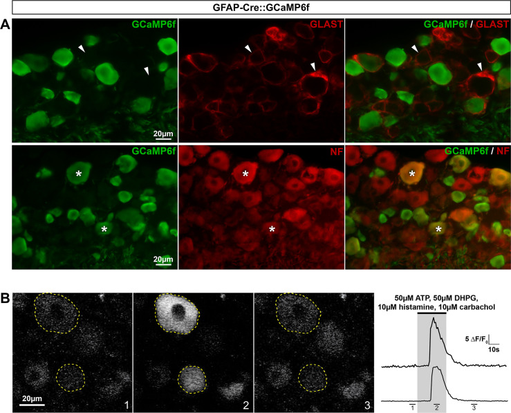
Fig 8. Cellular expression and functionality of GCaMP6f in GFAP-Cre::GCaMP6f mouse DRGs.
A, Representative images of immunohistochemistry in DRGs from GFAP-Cre::GCaMP6f mice showing GCaMP6f staining (top & bottom left panels, green), GLAST-expressing SGCs (top middle panel, red, arrowheads), and small and large sensory neurons (bottom middle panel, red, asterisks). Top & bottom right panels show superimposed pictures. For each row, scale bar in left picture applies to middle and right corresponding pictures. B, Representative images of 2-photon Ca2+ imaging experiment in ex vivo DRGs where neuronal GCaMP6f-expressing cell bodies (outlined areas of interest, left panel) exhibit intracellular Ca2+ increases; ➀ baseline, ➁ Gq GPCR agonist cocktail (50μM ATP, 10μM Histamine, 10μM Carbachol and 50μM DHPG) application, and ➂ wash (right panel).

