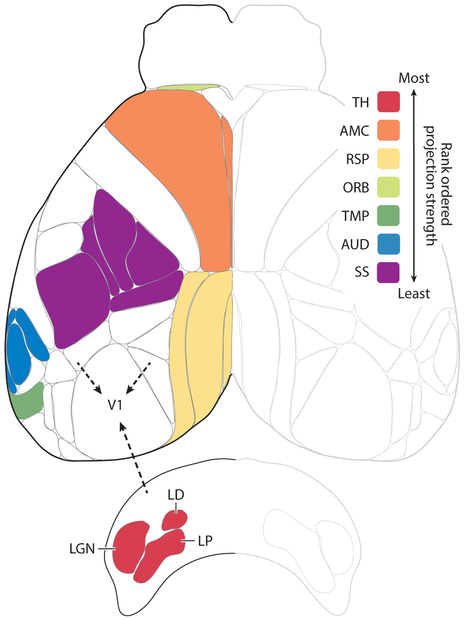
Figure 1.

Afferent projections to V1. Shown is a top view of the mouse neocortex based on the Allen common coordinate framework v.3 plus the thalamic nuclei that project to V1. Robustness of afferent projections outside of the visual cortex to V1 is shown rank ordered according to the density of projections adjusted for injection size. The inputs from most robust to least robust were: (1) the dorsal and ventral part of the lateral geniculate nucleus (LGN), lateral posterior (LP) and lateral dorsal nuclei (LD) of the thalamus (TH); (2) the secondary motor and anterior cingulate areas (AMC); (3) the retrosplenial areas and postsubiculum (RSP); (4) the orbital areas (ORB) (note that, for visibility reasons, only the area just anterior to AMC is indicated as part of the orbital areas); (5) the temporal, ectorhinal, perirhinal, entorhinal, postrhinal, subiculum, and CA1 areas (TMP) (note that only parts of the areas are visible); (6) the auditory areas (AUD); and (7) the somatosensory areas, including the whisker representation (SS). Details regarding the injections: We examined recombinant adeno-associated virus injections into wild-type mice from the Allen dataset into visual and non-visual areas and used a combination of projection pixel density and intensity (weighted by injection volume) to develop a qualitative ranking of projection strength (robustness) between areas. For V1 afferent connectivity, injections that also infected visual areas were discarded.
