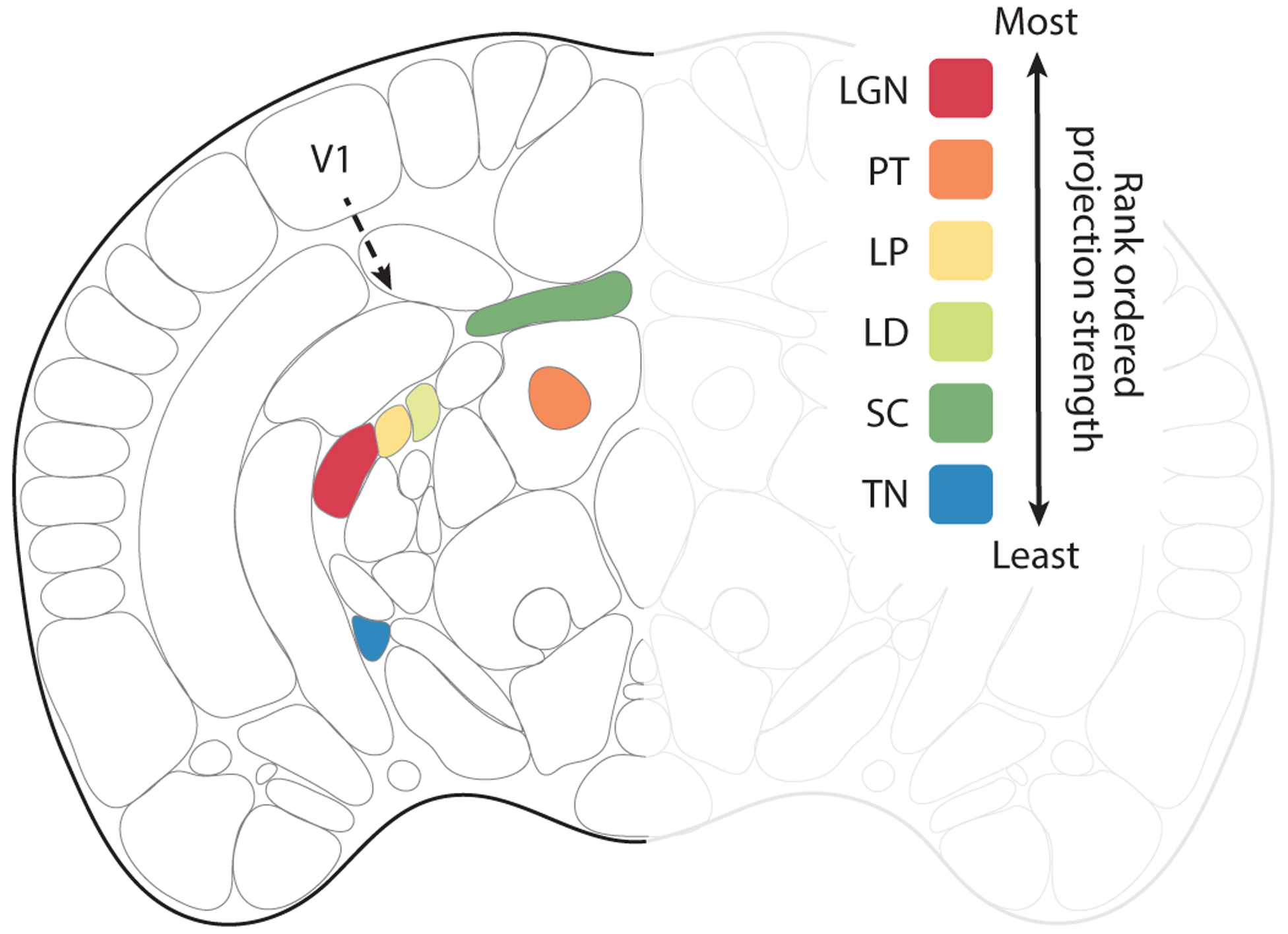
Figure 2.

Efferent subcortical projections from V1. Coronal view of the mouse brain. Based on the Allen common coordinate framework v.3 to include the lateral dorsal nucleus of the thalamus (LD) plus efferent projection zones of V1 (area 17). Robustness of efferent projections outside of the visual cortex is shown for the wild-type mouse, rank ordered according to the density of projections adjusted for injection size. The inputs from most robust to least robust were: (1) the lateral geniculate nucleus (LGN), (2) the pretectal nuclei (PT), (3) the lateral posterior nucleus of thalamus (LP), (4) the LD, (5) the superior colliculus (SC), and (6) the terminal nuclei (TN).
